Investigation of Regenerative Effects of Theranekron® and Misoprostol after Partial Hepatectomy in Rats
© 2023 Ismet Yilmaz, Burhan Hakan Kanat, Azibe Yildiz, Yilmaz Bilgiç, Ahmet Berk, et al., et al. This is an open-access article distributed under the terms of the Creative Commons Attribution License, which permits unrestricted use, distribution, and reproduction in any medium, provided the original author and source are credited.
Abstract
In the present study we were aimed to investigate/compare regenerative effects of Theranekron® (TC) and Misoprostol (MS) after Partial Hepatectomy (PH) in rats. This study was conducted in 38 rats, they were divided in to 5 groups and 7 days study periot. When considering biochemical and histopathological results; we were seen that after PH 7 days duration was not sufficient and also at least 10 days periot may be necessary. Althought some literatures were reported about positive effects of TC and MS on epithelial growing, we have not seen that they have not any positive effect on liver regeneration. In the future -at different doses and durations- these may be found effective.
Introduction
The liver has a crucial role in metabolism and regulation of blood glucose, protein synthesis, bile and urea production and all kind of detoxification. And also it maintains homeostasis and for this it acts in cooperation with many organs of body. After PH, regeneration process is regulated by certain growth factors, including HGF (hepatocyte growth factor), EGF (epidermal growth factor), TNF-α (tumor necrosis factor alpha), VEGF (vascular endothelial growth factor), TGF-β (transforming growth factor beta) and other cytokines [1].
Alcoholic extract of the spider Tarantula cubensis is licensed under the trade name TC. It is used in veterinary medicine in cases of septic conditions, such as dermatitis, inflammatory nail diseases (panaricium, foot rot), phlegumones, ulcers, abscesses, purulent lesions and necrosis. It is stated that it can be accelerate uterus involution and reduce vaginal discharge in the early postpartum period, and also it may be used as an alternative therapeutic for the treatment of clinical and subclinical mastitis in dairy cattle [2-4]. Additionally, in blue tongue infections of cattle, administration of TC together with oxytetracycline and flunixin have shown fever returns to normal earlier and re-epithelization accelerates, and it is more effective than alone levamisole administration in the treatment of nipple papillomatosis [5,6]. It is stated that TCapplication as an additional treatment in the treatment of tongue erosion in cats completely passes erosion and re-epithelialization is observed in the upper lip ulcer [7]. It has been stated that, in the endometriasis model, TC application prevented relapses accelerated wound healing after laparotomy and may be partially prevent oxidative stress after exposure to aflatoxin in rats [8-10].
MS is a prostaglandin E1 analogue and an approved drug against non- steroidal inflammatory gastric ulcers and is also used offlabel in the clinic to induce labor pain [11]. It has been stated that MS has a protective effect against mucosal lesions in the stomach and liver damage patients with hepatitis B [12]. In mice a lipopolysaccharide induced endotoxicity model, MS had positive effects on brain antioxidant parameters and decreased serum aspartate transaminase (AST) and alanine transaminase (ALT) levels and in rats it has been reported to have a protective effect against acetaminophen- induced liver damage and cisplatininduced ototoxicity, same animals it has a protective effect against acetaminophen-induced microvascular liver damage, and a positive effect against CCl 4 - induced liver damage [13-17]. Some researchers have been reported that, prostaglandins (PGs) (as to be cytoprotective) are triggering the liver regeneration cascade by using proliferative factor (include HGF, EGF, and interleukin-6 (IL- 6)) after PH in rats [18]. It has been reported that, in mammals the regenerative capacity of the remnant liver changes depending on genetics, nutritional status and the food and/or drugs used, and also in rats regeneration process is completed within 7-10 days after PH [19, 20].
In literature reviews, there was not found any study report about the regenerative effects of TC and MS. Although TC is licensed in veterinary practice for use in the treatment of the some animal diseases mentioned above, our aim is to investigate its possible benefits such as liver regeneration in human medicine and we believe that this will be the most original part of our work. Additionally, given information above about these two drugs and our previous experimental studies on liver regeneration have led us to comparison of their regenerative effects in rats. In this study; comparing the regenerative effects of MS and TC after partial hepatectomy have examined separately and combined (synergism/antagonism) and findings have been supported by histological and biochemical datas in rats.
Material and Methods
Animals and Study Protocol
In this study, 38 Sprague Dawley female rats were used. Rats were obtained from the Experimental Animal Reproducing and Investigational Center of Inonu University. The principles of the Ethics Board of Inonu University, Faculty of Medicine were followed throughout the study (Ethics Board Protocol No:2018/ A-23). The rats were 15-16 months years old and their avarage body weights were measured as 314,8±41,5 g and they were maintained in standard cages until the the experiment. The rats were kept in rooms equipped with 12 h, dark-light lighting cycles, housed four per cage under appropriate humidity (54-56%) and ventilation conditions at room temperatures between 25±2 °C. From the beginning to the end of the study they were fed normal rat chow and tap water ad libitum
The animals were randomly divided into five groups as follows
• Group (Control-C) (n=6): After study beginning of 7 days
the rats were under only laparatomy, and after laparatomy
on 7th days they were humane killing.
• Group (Sham-S) (n=8): After study beginning of 7 days
the rats were under 70% PH, and after PH on 7th days they
were humane killing.
• Group (MS) (n=8): At the beginning of study, 0,1 mg/kg/
day MS were given by oral route, and after 7 days, 70% PH
were done, and at PH on 7th days they were humane killing.
• Group (TC) (n=8): At the beginning of study, 0,2 mg/kg/day
TC were administred by s.c. route, and after 7 days 70%PH
were done, and after PH on 7th days they were humane killing.
• Group (MS+TC) (n=8): At the beginning of study, 0,2 mg/
kg/day TC were administred by s.c. route + 0,1 mg/kg/day MS
were given by oral route, and after 7 days 70% PH were done,
and after PH on 7th days they were humane killing. Taken
blood samples (app. 8 mL) and liver tissue samples were used
for biochemical and histopathological examinations. After
PH, within 24 hours totally 4 rats were died for undetermined
cause (2 rats in group 4 and 2 rats in group 5).
In this study, Theranekron®; D6, 50 mL (Serial number:1118769AA, Expirate data; 10.2021 Richter pharma AG, Wels, Austria) and Misoprostol 10mg, Santa Cruz Biotechonology, USA (Lot K; 1219,) were used. At the beginning of study (first seven days), the TC was administered s.c. route as 1 mL TC+15 mL SF, (1mL/16 rats=0,2 mg/kg/day, totally 16 rats=~5 Kgr), after PH (second seven days), the dose adjustment is required due to deaths, 0,8mL TC+11,2mLSF, (1mL/12 rats, totally 12 rats=~4 Kgr).
Phosphate Buffered Saline (PBS) tablet was dissolved in 100 mL distilled water and this solution was prepared. At the beginning 10 mg of MS was attempted to be dissolved in 6 ml of PBS, but a clear solution was not formed. Subsequently, the PBS amount was increased to 10 ml and the stock solution was prepared. The daily dose required for 16 rats (totally 16 rats=~5 kg) to be treated with MS was calculated and as to be 0,5 mg / kg / day. And 0.5 ml each of the stock solution was taken into eppendorf tubes and stored at -20 °C. Every day this eppendorf tubes were held on 1 hours at room temperature and 0.5 mL of MS stock solution + 15.5 mL of PBS was taken in to 20 mL enjectors and administered with gavage by oral route to be as 1 mL / rat dose.
Surgical Procedure
All surgical procedures (Laparotomy and 70% PH) were performed under sterile conditions. Rats were anesthetised by intraperitonal injection of ketamine (50 mg/kg) and xylazine (10 mg/kg) as anesthetic agents, PH was performed according to the method of Higgins and Anderson [21]. In the supine position, an upper midline incision of the abdomen was followed by retraction of the xyphoid cartilage for adequate exposure of the liver and division of hepatic ligaments. The right median, left lateral, and median liver lobes (anterior lobes), which correspond to approximately 70% of the total liver mass were resected, whereas the right lateral and caudate lobes (posterior lobes) were left intact. After irrigation of the abdomen with warm saline, the peritoneum and skin were closed with running 4-0 and 3-0 sutures, respectively. Postoperatively, animals were allowed to recover from anesthesia with free access to food and water until humane killing. For sham surgeries, livers were externalized and gently palpated to mimic the surgical stress of the PH procedure. Animals undergoing PH did not receive any antibacterial agents. On 7th days postoperative, all animals were anesthetized by intraperitoneal injection of ketamine + xylazine for humane killing.
Biochemical Evaluation
Blood samples for serum biochemical analysis were left to stand for half an hour, it was subsequently centrifuged (at 3,500 rpm for 10 min) and obtained serum samples were used to assess liver function parameters like AST, ALT, alkaline phosphatase (ALP), gamma-glutamyl transferase (GGT), cholesterol (mg/dL) andtotal bilirubin (mg/dL). All serum parameters were measured by AutoAnalyzer (Beckman Coulter AU5800, Inonu University, Turgut özal Medical Center Biochemistry Laboratory). The serum liver enzymes were expressed as U/L.
Tissues were dissected and washed immediately with cold PBS (pH 7.4), dried and weighed. Ten volumes of PBS were added per gram of tissues and were homogenized (IKA Ultra Turrax). Homogenates were centrifuged at 20,000 g for 15 minutes (Eppendorf 5415R). Supernatants were separated and kept at -80 °C until all measurements were performed. The protein content of the tissues was measured according to the Bradford method using bovine serum albumin as the standard [22]. Results were expressed as milligram protein. For the determination of antioxidant and lipid peroxidation, superoxide dismutase (SOD) and catalase (CAT), glutathione (GSH), and malondialdehit (MDA), a marker of lipid peroxidation were analyzed using commercial ELISA kits from Bioassay Technology Laboratory, Cat. No E1101Ra (GSH), E0869Ra (CAT), 0156Ra (MDA), 0168Ra (SOD) and expirate date: May 2021.
Histopathological Analysis
The liver was fixed in 10% neutral formaldehyde. After fixation, specimens were dehydrated in 70%, 80%, 90%, 96% and 99% alcohol series. In the next step, the tissue specimens were cleared in xylene, and then embedded in paraffin. Paraffin-embedded specimens were cut into 4 μm thick sections, mounted on slides. The tissue sections were stained with hematoxylin- eosin (H-E) for general liver structure. The liver damage was semiquantitatively assessed as follows; sinusoidal congestion and dilatation, inflammatuar cell infiltration and hepatocytes degeneration (eosinophilic cytoplasm, pyknotic nuclei, vacuolation). For each criterion, ten fields were examined in 20x objective magnification. Accordingly, the extensiveness of liver damage were scored between 0-3; 0 was defined as normal liver, 1 was defined as liver damage involving <25% of liver, 2 was defined as liver damage involving 25% - 50% of liver and 3 was defined as liver damage involving >50% of liver. Leica DFC280 light microscope and a Leica Q Win Image Analysis system (Leica Micros Imaging Solutions Ltd., Cambridge, UK) was used for all histopathological and immunohistochemical evaluations and measurements.
Immunohistochemical Analysis
To assess hepatocyte proliferation, immunostaining was performed with PCNA and Ki-67 antibody. PCNA, which is a cofactor of DNA polymerase, was prevalent in the nuclei of proliferating hepatocytes. Ki-67, a specific marker of proliferation, and it was also present in the nuclei of proliferating cells. For immunohistochemical analysis, 4 μm thickness tissue sections were deparaffinized and rehydrated and placed in antigen retrieval solution (citrate buffer, pH 6.0) and boiled in a pressure cooker for 20 minutes and cooled to room temperature for 20 min. Then the sections were washed with PBS (pH 7.4), then for blocking endogenous peroxidase activity the slides incubated in 3% hydrogen peroxide solution for 15 min at room temperature and were washed in PBS. After the blocking of non-specific antigenbinding sites with protein block, Ki-67 (ThermoFisher Scientific, Fremont, USA) and PCNA (Santa Cruz Biotechnology, Inc., Heidelberg, Germany) antibodies were applied for 60 minutes at room temperature. After being rinsed with PBS, sections were incubated with biotinylated secondary antibody and streptavidin peroxidase for 10 minutes at room temperature. Samples was visualized with the chromogenic substrates AEC, counterstained with hematoxylin, mounted in glass slide. The nucleus of Ki-67 and PCNA positive cells stained as brown color. After Ki-67 and PCNA positive hepatocytes were counted in 10 randomly chosen high-power (x400) fields from three sections per group, means were calculated for analysis (Table-3).
Statistical Analysis of Histopathology and Immunohistochemictry Data was analyzed using the IBM SPSS software program for Windows, version 22.0 (SPSS Inc., Chicago, IL). The data was expressed as either medians or means ± SD depending on the overall variable distribution. The normality of the distribution was confirmed using the Shapiro-Wilk test. The normally distributed data was analyzed by one-way ANOVA followed by the Tukey post hoc test. The non-normally distributed data was compared by the Kruskal-Wallis H test. When significant differences were determined, multiple comparisons were carried out using the Mann- Whitney U test with Bonfernoni correction. Results were considered statistically significant at p<0.05.
Results
A. The Liver Weights: In the present study, we were recorded resected (during PH operation) and totally (at study finish) liver weights, proportionally there were not any significant differences our before study results [19].
B. The Serum Liver Parameters: Effects of TC and MS on serum biochemical parameters in control, sham, and treated groups were summarized in table 1. Levels of total bilirubin in TC and MS treated groups were not changed compared to the normal control groups. While there was a significant decrease in level of cholesterol in the MS and MS+TC groups compared to the control, there was no significant difference in sham and treated groups. The hepatic function has been monitored by the evaluation of the serum level of transaminases such as ALT and AST. Also liver enzymes, ALP and GGT were analyzed. Table 1 illustrates the effect of TC and MS on liver marker enzymes in control, sham and treated groups. Serum ALT levels were increased in the sham group compared to the control. In the treatment groups a significant decrease was found in the TC group. We observed that the serum AST levels increased in the sham group compared to the control. Among the treatment groups a significant decrease was found in the TC group compared to the control and sham groups. We also found a significant decrease in the MS+TC group compared to the sham group. Although serum ALP levels increased in the sham group compared to the control group a non-significant decrease was observed in the TC group compared to the control and sham groups. In addition there was a significant increase in the MS+TC group compared to the control and sham groups. Serum GGT levels increased in the sham group compared to the control group and a significant decrease was observed in the TC group compared to the sham group.
C. Liver Tissue Oxidative Stress Parameters: For evaluation of effects of TC and MS on liver oxidative stress status (SOD, CAT, GSH, and MDA levels) was analyzed. Results were presented in table 2 and figure 1. While there was a significant decrease in the SOD level in the sham group compared to the control, a nonsignificant increase was found in all treatment groups compared to the sham group. The MDA level in the sham group increased significantly compared to the control and a non-significant decrease was obtained in all treatment groups compared to the sham group. CAT and GSH levels were not comparable. The serum biochemical analysis: ALT, AST, ALP, GGT, cholesterol and total bilirubin levels were presented in table 1.
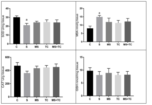
Figure 1: Liver tissue SOD, MDA, CAT and GSH levels at 7th days of PH in rats
D. Statistical Analysis of Serum Liver Enzymes and Tissue Oxidative Stress Parameters: Data were given as median (min-max). Kruskal Wallis test was used to determine the statistically difference between groups. Conover test was used in multiple comparisons after the Kruskal Wallis test. A p value of <0.05 was considered statistically significant. In the analyses, “Statistical Analysis Software”; was used, this programme was developed by Inonu University Biostatistics and Medical Informatics Department [23].
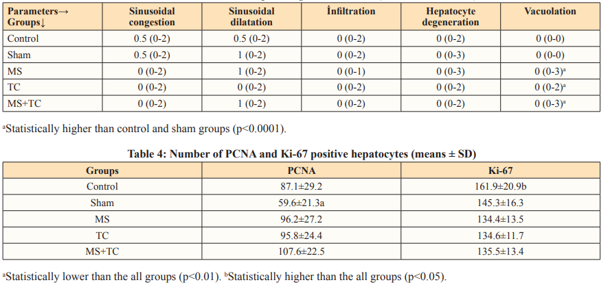
E. Histopathological Results: The sections stained with H-E staining method, sham group showed a normal appearance of the liver histology except minimal changes (Fig. 2A). The control group exhibited similar histological appearance to the sham group (Fig. 2B). On the other hand, vacuolization was observed in hepatocytes in the MS, TC and MS+TC groups different from the control and sham groups (Fig 2C, D, and E). The MS, TC and MS+TC groups was statistically different from the sham and control groups in terms of the presence of vacuolization (p<0.0001). The histopathological evaluation results are given in Table 3.
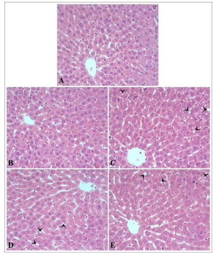
Figure 2: The appearance of the liver in the sham (A), control (B), MS (C), TC (D) and MS+TC (E) groups. The presence of vacuolization (arrow heads) in hepatocytes in the MS, TC and MS+TC groups is noticeable. H-E, x40.

Figure 3: The PCNA positive hepatocytes (arrow heads) in the sham (A), control (B), MS (C), TC (D) and MS+TC (E) groups. PCNA immunostaining x40.
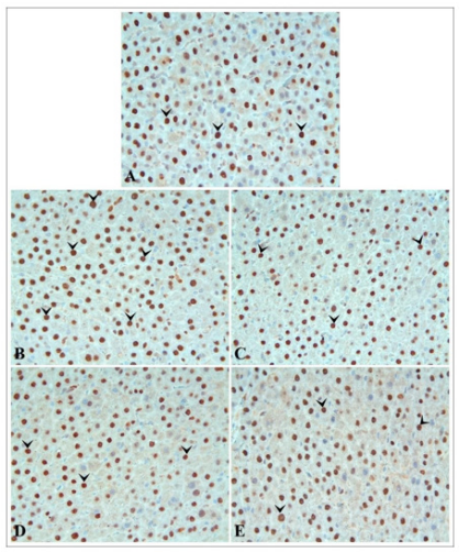
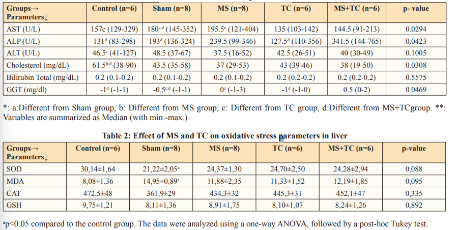
Figure 4: The Ki-67 positive hepatocytes (arrow heads) in the sham (A), control (B), MS (C), TC (D) and MS+TC (E) groups. Ki67 immunostaining x40.
F. Immunohistochemistry: The proliferation of hepatocytes was estimated using immunohistochemical staining for the PCNA and Ki-67. Hepatocyte nuclei showing a positive reaction by the PCNA and Ki-67 immunostaining method were stained brown (Fig. 2 and 3). Number of PCNA positive cells were detected significantly increase in the control, MS, TC and MS+TC groups compared to sham group (p<0.01). On the other hand, the number of cells in these groups was found similar. Number of Ki-67 positive cells were significantly increased in the control group compared to all other groups (p<0.05). On the other hand, number of Ki-67 labeled nuclei was statistically similar in the sham, MS, TC and MS+TC groups. Number of PCNA and Ki-67 positive hepatocytes were given in Table 4.
Discussion
In the present study, dose of MS was same with Salam et al 2009 and Bilgiç & Ozgocmen, and ten folder lower from Smith and Lautt. The dose of TC was three times lower than Kizilay and Adib Hashemi et al [24-26]. Probably, if the doses of MS and TC were used twice or three times higher than used in this study the results could be more meaningful.
In the present study, our AST levels were higher than our previous reports, the ALT levels were shown important differences from our previous reports [18-19]. And also ALP levels were shown similarities with these reports (Table 1). The SOD levels of this study were shown significantly differences from our reports, especially from our the first one [18-19]. The CAT levels of this study were shown similarities with the firts ones, but this similarities were not shown with other (Table 2).
Table 1: The serum liver parameters
The histopathological evaluation results (Table 3) of present study was similar to our study [18]. But PCNA results (Table 4) of present study were significantly different from our previous reports [19]. These differences may be due to from measurement methods, from the sensitivity of the device, preparation of samples, used animal age and sex differences. In histopathological examinations; the presence of vacuolization has a significant value for cell life and death. Sharma et al were reported that, depending on the dose and time sorafenib treatment increases reactive oxygen species (ROS) levels in cells along with cytoplasmic vacuolation due to endoplasmic reticulum (ER) luminal dilation that are independent of autophagy [24-27]. These findings (either TC or MS) may have an importance to future studies and it must be considering especially in the farm animal treatments.
Table 3: The results of histopathological evaluation (med with min-max)
When liver cell membrane is damaged, a variety of enzymes located normally in cytosol is released into the blood, thereby causing increased enzyme level in the serum [28,29]. For estimation of hepatocellular damage, enzymes in the serum are used as a useful quantitative marker. In the present study, we measured aminotransferases (ALT, AST) and GGT enzymes, cholesterol, and total bilirubin levels for assessing liver function. Serum transaminases and ALP levels are considered to be the most sensitive markers [30]. ALT is more liver specific to determine hepatocellular damage [31]. It is reported that these enzymes were increased during partial hepatectomy [32-34]. In our study, although there was an increase in ALT, AST, ALP and GGT enzymes in the hepatectomy group compared to the control group, but there was not any statistically significant. In the treatment groups, the amount of ALT was significantly reduced by TC treatment. Although other enzyme levels also decreased in the TC group, this result was not significant. It is reported that liver damage ameliorated 72 hours after resection [35]. So our results may have occurred because that we measured our samples on the 7th day after liver resection.
Serum bilirubin is considered as one of the test of liver functions. Bilirubin is produced after Hem catabolism and conjugates with glucuronic acid in hepatocytes for excretion in to the bile [36]. Bilirubin levels were not comparable between the all groups in the present study. To evaluate the extent of liver injury serum GGT were analysed. We observed that GGT levels increased after PH and decreased significantly with T treatment. A positive effect of the other treatment groups was not observed. The liver shows a notable regenerative capacity after PH and free radicals appear during PH [37]. Because of that oxidative stress plays a role in PH, antioxidant agents may be beneficial for regeneration after PH [38]. Many studies have presented an increased production of free radicals such as MDA in liver mitochondria following partial hepatectomy. The activities of antioxidant enzymes protect cells against oxidative damage [39]. In our study, we observed a significantly increased MDA and in treatment groups, MDA levels decreased none significantly. We couldn’t find a statistically significant difference between PH and treatment groups. GSH levels were not comparable between the groups. Also SOD and CAT enzymes were measured as indices of the antioxidant status of liver tissues. SOD catalyzes the dismutation of the superoxide radical to hydrogen peroxide. While a statistically significant decrease was observed after PH, SOD levels increased in the treated groups, but this increase was not significant. No changes were observed in the antioxidant enzymes between PH and treatment groups. CAT is an antioxidant enzyme that degrades endogenously produced hydrogen peroxide [40]. CAT activity is reduced in hepatic injury [41]. After PH there was a decrease in CAT compared to the control levels, but it was not significantly. In treatment groups, CAT levels increased, they were not significantly too.
Consequently: before the present study, we were published two study reported on about liver regeneration [19-20]. In these reports for regeneration we were considered 21 and 10 days after PH, but in the present study we were seen that 7 days duration was not sufficient, and also at least 10 days periot may be necessary. Althought some literatures were reported that, TC has been found an effective agent on the treatments of some animal diseases we have not seen any positive effect on liver regeneration, and also same situation may have say for MS. But, in the future (at different dose and durations) these may be found effective [2-8].
Competing Interests
The authors have declared that no competing interests exist.
Acknowledgments
This study was supported by the Scientific Research Fund of Inonu University (Project number: TSA-1778). The authors would like to express profound thanks for the financial support to the Unit of Scientific Research Projects of Inonu University, Malatya, Türkiye.
Ethical Steatment
The principles of the Ethics Board of Inonu University, Faculty of Medicine were followed throughout the study (Ethics Board Protocol No:2018/ A-23).
References
- Lee GS, Yang HG, Kim JH, Ahn YM, Han MD, et al. (2019) (Pinus densiflora) needle extract could promote the expression of PCNA and Ki-67 after partial hepatectomy in rat. Acta Cir Bras 34:
- Kaçar C, Kaya S (2014) Uterine infections in cows and effect on reproductive performance. Kafkas Univ Vet Fak Derg 20:
- Lotfollahzadeh S, Alizadeh MR, Mohri M, Mokhber Dezfouli MR (2012) The therapeutic effect of Tarentula cubensis extract (Theranekron®;) in foot-and-mouth disease in cattle: a randomised trial in an endemic setting. Homeopathy 101:
- Gürbulak K, Akçay A, GümüŞsoy KS, Sist B, Steiner S, et al. (2014) Investigation of the efficacy of Tarantula cubensis extract (Theranekron D6) in the treatment of subclinical and clinical mastitis in dairy cows. Turk J Vet Anim Sci 38:
- Albay Mk, Sahinduran S, Kale M, Karakurum Mc, Sezer K (2010) Influence of Tarantula cubensis extract on the treatment of the oral lesions in cattle with bluetongue disease. Kafkas Univ Vet Fak Derg 16:
- Paksoy Z, Gülesci N, Kandemir FM, Dinçel GC (2015) Effectiveness of Levamisole and Tarantula Cubensis extract in the treatment of teat papillomatosis of cows, Indian J Anim Res 49:
- Yardimci C, Yardimci B (2008) Indolent ulcer in a cat. Ankara üniv Vet Fak Derg 55:
- Dolapcioglu K, Dogruer G, Ozsoy S, Ergun Y, Ciftci S, et al. (2013) Theranekron for treatment of endometriosis in a rat model compared with medroxyprogesteroneacetate and leuprolideacetate. Eur J Obstet Gynecol Reprod Biol170:
- Adib-Hashemi F, Farahmand F, Hesari SF, Rezakhaniha B, Fallah E, et al. (2015) Anti-inflammatory and protective investigations on the effects of Theranekron(R) “an alcoholic extract of the Tarantula cubensis”; on wound healing of peritoneal in the rat: an in vivo comparative study. Diagn Pathol 10: 10-19 10. Karabacak M, Eraslan G, Kanbur M, Sarica ZS (2015) Effects of Tarantula cubensis D6 on aflatoxin-induced injury in biochemical parameters in rats. Homeopathy 104:
- Chen W, Xue J, Peprah MK, Wen SW, Walker M, et al. (2016) A systematic review and network meta-analysis comparing the use of Foleycatheters, misoprostol, and dinoprostone for cervical ripening in theinduction of labour. BJOG 123:
- Flisiak R, Prokopowicz D (1997) Effect of misoprostol on thecourse of viral hepatitis B. Hepato gastroenterology 44:
- Abdel-Salam OME, Mohammed NA, Morsy SMY, Youness ER, Omara EA, et al. (2014) Misoprostol decreases oxidative stress and liverinjury in bacterial lipopolysaccharide-induced endotoxemia in mice. Comparative Clin Pathol 23:
- Lim SP, Andrews FJ, O’Brien PE (1994) Misoprostol protection against acetaminophen-induced hepatotoxicity in the rat. Dig Dis Sci
- Dogan M, Polat H, YaŞar M, Kaya A, Bayram A, et al. (2016) Protective role of misoprostol against cisplatin-induced ototoxicity. Euro Arch Oto-Rhino-Laryngol 273:
- Lim SP, Andrews FJ, O’Brien PE (1995) Acetaminopheninduced microvascular injury in therat liver: protection with misoprostol. Hepatology 22:
- Salam OM, Sleem AA, Omara EA, Hassan NS (2009) Hepatoprotective effects of misoprostol and silymarin on carbontetrachloride-induced hepatic damage in rats. Fundam Clin Pharmacol
- Smith JMS, Lautt WW (2005) The role of prostaglandins in triggering the liver regeneration cascade. Nitric Oxide 13:
- Yilmaz I, Hatipoglu HS, TaŞlidere E, Karaaslan M (2018) Comparing the regenerative effects of silymarin and apricot on liver regeneration after partial hepatectomy in rats. Biomed Res 29:
- Yilmaz I, Karaman A, Vardi N, çetin A, Erdemli E (2013) Effects of organic apricot on liver regeneration after partial hepatectomy in rats. Transplant Proceed 45:
- Higgins GM, Anderson AR (1931) Experimental pathology of the liver. Restoration of the liver of the white rat following partial surgical removal. Arch Pathol 12:
- Bradford MM (2020) A rapid and sensitive method for the quantitation of microgram quantities of protein utilizing the principle of protein-dye binding. Anal Biochem 72:
- YaŞar Ş, Arslan A, Colak C, Yologlu S (2020) A Developed Interactive Web Application for Statistical Analysis: Statistical Analysis Software. Middle Black Sea J Health Sci 6:
- Bilgiç S, özgöçmen M (2019) The protective effect of misoprostol against doxorubicin induced liver injury. Biotec Histochem 94:
- Kizilay Z, Aktas S, Kahraman Cetin N, Kiliç MA, Oztürk H (2019) Effect of Tarantula Cubensis Extract (Theranekron) on Peripheral Nerve Healing in an Experimental Sciatic Nerve Injury Model in Rats. Turk Neurosur 29:
- Adib-Hashemi F, Farahmand F, Hesari SF, Rezakhaniha B, Fallah E, et al. (2015) Anti-inflammatory and protective investigations on the effects of Theranekron®; ”;an alcoholic extract of the Tarantula cubensis”; on wound healing of peritoneal in the rat: an in vivo comparative study. Diag Pathol 10:
- Sharma S, Ghufran SM, Ghose S, Biswas S (2021) Cytoplasmic vacuolation with endoplasmic reticulum stress directs sorafenib induced non apoptotic cell death in hepatic stellate cells. Nature portfolio Scientific Reports 11:
- Kawaguchi D, Hiroshima Y, Kumamoto T, Mori R, Matsuyama R, et al. (2019) Effect of portal vein ligation plus venous congestion on liver regeneration in rats. Annals Hepatol 18:
- Rajesh MG, Latha MS (2004) Preliminary evaluation of the antihepatotoxic effect of Kamilari, a polyherbal J Ethnopharmacol 91:
- Sturgill MG, Lambert GH (1997) Xenobiotic-induced hepatotoxicity: mechanisms of liver injury and methods of monitoring hepatic function, Clin Chem 43:
- Lin CC, Shieh DE, Yen MH (1997) Hepatoprotective effect of the fractions of Ban-zhi-lian on experiment liver injuries in rat. J Ethnopharmacol 56:
- Yao H, Fu X, Zi X, Jia W, Qiu Y (2018) Perioperative oral supplementation with fish oil promotes liver regeneration following partial hepatectomy in mice via AMPK Mol Med Rep 17:
- Barros PP, Silva GH, Gonçalves GMS, Oliveira JC, Pagnan LG, et al. (2017) Hepatoprotective Effect of Quercetin Pretreatment against Paracetamol-Induced Liver Damage and Partial Hepatectomy in Rats. Braz Arch Biol Technol 60:
- He Z, Lou K, Zhao J, Zhang M, Zhang L, et al. (2020) Resina Draconis Reduces Acute Liver Injury and Promotes Liver Regeneration after 2/3 Partial Hepatectomy in Evidence-Based Complem Alter Med. 2020:
- Furuta K, Kakita A, Takahashi T, Tomiya T, Fujiwara K (2000)Experimental study on liver regeneration after simultaneous partial hepatectomy and pancreatectomy. Hepatol Res 17:
- SF, Martin P, Munoz JS (2003) Laboratory evaluation of the patient with liver disease. Hepatology, a textbook of liver disease. Philedelphia; Saunders publication 1:
- Fausto N, Campbell JS, Riehle KJ (2006) Liver Hepatology 43:
- Dayoub R, Vogel A, Schuett J, Lupke M, Spieker SP, et (2013) Nrf2 activates augmenter of liver regeneration (ALR) via antioxidant response element and links oxidative stress to liver regeneration. Mol Med 19:
- Aguilar-Delfin I, Lopez-Barrera F, Hernandez-Munoz R (1996) Selective enhancement of lipid peroxidation in plasma membrane in two experimental models of liver regeneration: partial hepatectomy and acute CC14 Hepatology 24:
- Nicholls P, Fita I, Loewen PC (2000) Enzymology and Structure of Catalases. Advances Inor Chem. 51:
- Atmaca M, Bilgin HM, Obay BD, Diken H, Kelle M, et (2011) The hepatoprotective effect of coumarin and coumarin derivates on carbon tetrachloride-induced hepatic injury by antioxidative activities in rats. J Physiol Biochem 67: 569- 576.