Hyper-Stimulation of the Endometrial Thickness and Egg Relaese of Women between the Ages of 18-46 years in Imo State, Nigeria
© 2024 Emeonye Odochi Peace, Obiyo Franklin C, Okoroafor Chimezie, AGU Ifenyinwa S, Ukagu Ngozi C, et al. This is an open-access article distributed under the terms of the Creative Commons Attribution License, which permits unrestricted use, distribution, and reproduction in any medium, provided the original author and source are credited.
Abstract
ABSTRACT
The uterus is a dynamic tissue under the control of ovarian steroids, estrogen and progesterone, it comprises of endometrial layer of the uterus which undergoes proliferation, secretion and menstrual cycle to reach the receptive state in the implantation of the embryo. Endometrial thickness and growth during ovarian stimulation is a possible predator of implantation in invitro fertilization.
Objective: To ascertain the endometrial thickness and number of ovulation egg released by hyper-stimulation. Research Design: The study population contains 10 women aged 18-46 years. Groups were randomly selected from clients that came to seek for medical help in vaden specialist clinic, Owerri, Imo State Nigeria.
Result: The age group 18-25 had the highest thickness of endometrium (45MM thick) while the age group 36-40 had the least thickness of endometrium (7MM thick). The age group 20-25 produced the greatest quantity of eggs (53.8%) while the age group 36-40 produced the least quantity of eggs (0.77%).
Summary: This study has shown that Endometrial thickness increases under hormonal influence which affect the egg release from the ovaries.
Introduction
The Endometrium is the inner mucous membrane lining the uterus. It is further subdivided into 2 parts either Deep stratum basalis: This change little throughout the menstrual cycle and is not shed at menstruation. Superficial stratum functionalis: Which Proliferates in response to oestrogens, and becomes secretory in response to progesterone. It is shed during menstruation and regenerates from cells in the stratum basalis layer. The endometrium layer of the uterus undergoes proliferation, secretion and menstrual cycle to reach the receptive state in the implantation of the embryo. When the endometrial carries out its natural functions by stimulation of the hormones, the effects of this stimulation arises as transient secretion of endometrial proteins, changes in cell behavior and thickness. Endometrial thickness and growth during ovarian stimulation is a possible predator of implantation in invitro fertilization.
As women age, they experience a decline in reproductive performance leading to menopause. This decline is tied to a decline in the number of ovarian follicles. Although about 1 million oocytes are present at birth in the human ovary, only about 500 (about 0.05%) of these ovulate, and the rest are wasted. The decline in ovarian reserve appears to occur at a constantly increasing rate with age, and leads to nearly complete exhaustion of the reserve by about age 52 [1]. As ovarian reserve and fertility decline with age, there is also a parallel increase in pregnancy failure and meiotic errors resulting in chromosomally abnormal conceptions. The ovarian reserve and fertility perform optimally around 20–30 years of age. Around 45 years of age, the menstrual cycle begins to change and the follicle pool decreases significantly. The events that lead to ovarian aging remain unclear.
Research Methodology
The study population contains 10 women aged 18-46 years. Groups were randomly selected from patients that came to seek for medical help in vaden specialist clinic, Owerri, Imo State Nigeria.
Study Setting
This is a population study conducted on 10 individuals ranging from 18-46years that came to Vaden Specialist hospital Owerri.
Ethical Clearance Ethics
Because the study involved morality ethical consent was sorted from the individuals and their co-operation was remarkable and many of them formally permitted me to do the study on them. Inclusion criteria: for individual that have 28day menstrual cycle who have recorded 3 consecutive menses. The individuals were warned to abstain from coitus during this period.
Procedure
To ascertain the endometrial thickness, the following instruments were used to visualize the uterus and the ovarian status Ultrasound Machine, Ovulation strip and Laparoscopy.
Study Method
The method involved applies injecting the drug becinolyn into the individual at mid menstrual cycle which corresponds to the day of ovulation at day 14th from the 1st day, for individuals with 28day menstrual cycle. There after another drug called menopor at 225iu was injected in the individual on her 1st day of menstrual cycle which is day 28 often the first day of last menstruation. Thirteen days were allowed for the drug action to run its full course, after which follicles were harvested from the two ovaries of the individual by Ultrasound- Guided trans vaginal eocytes vetrieval method, (gold standard) for human in vitro fertilization (IVF) [2-17].
Results
Table 1: Age Distribution of the 4.1 Study Population “n”=10
|
Age range in years |
Frequency |
Percentage (%) |
|
20-25 |
4 |
50 |
|
26-30 |
2 |
25 |
|
31-35 |
- |
- |
|
36-40 |
2 |
12.5 |
|
41-46 |
2 |
12.5 |
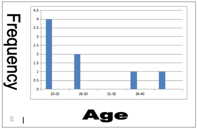
Figure 1: Age Distribution of the Population “n”=10
The Above table showed the frequency of the age 20-25 (4) representing 50%, 26-30 (2) representing 2%. while 36 to 46 (25) represent 25% of the population.
Table 2: The Normal Endometrium Thickness in a Normal State is 3MM Showed the Drugs used in Stimulation of the Egg Release and Endometrial Thickness
|
S/N |
CLIENTS |
AGE |
DRUG DOSAGE MENOPUR |
DURATION |
NO OF EGGS RELEASED |
ENDOMETRIAL THICKNESS |
|
01 |
A |
20yrs |
225IU |
13days |
17eggs |
10.MM |
|
02 |
B |
25yrs |
225IU |
13days |
24eggs |
15MM |
|
03 |
C |
29yrs |
225IU |
13days |
17eggs |
10MM |
|
04 |
D |
20yrs |
225IU |
13days |
10eggs |
9MM |
|
05 |
E |
21 yrs |
225IU |
13days |
19 eggs |
10MM |
|
06 |
F |
29yrs |
225IU |
13days |
3 eggs |
7MM |
|
07 |
G |
42yrs |
225IU |
13days |
12eggs |
9MM |
|
08 |
H |
38 Yrs |
225IU |
13days |
1egg |
6MM |
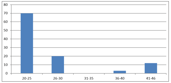
Figure 2: Showed the Drugs Used in Stimulation of the Egg Release and Endometrial Thickness
The Above Table Showed that the Dosage of The Drug Used to Stimulate Ovulation Was the Same in All the Ages Including the Study Days (13). With the age Group (20-25yrs) Having the Highest Frequency; These Findings Show that Frequency of Egg Produced and Endometrial Thickness is Predominated in the Age Bracket (20-25) Years Whereas Low Frequency of Egg Production and Endometrial Thickness Frequency Was Seen in Ages (36-40) and (41-46) Years. This Is Because Egg Production Is Subject to Hormonal Stimulation Which Increases Naturally From (20-25yrs) Old but Becomes Low as The Individual Ages [2].
Table 3: Endometrial Thickness in the Study Population After hyperstimulating the ovary using 225IU of menopur and becinolyn for 13days, the thickness of the endometrium was accessed using ultrasound machine and recorded separately for the different age groups.
|
Age Range in Years |
Endometrial Thickness |
|
20-25 |
45MM |
|
26-30 |
17MM |
|
31-35 |
0MM |
|
36-40 |
7MM |
|
41-46 |
10MM |
|
Total |
79MM |
The age group 20-25 had the highest thickness of endometrium (45MM thick) while the age group 36-40 had the least thickness of endometrium (7MM thick).
This is futher highlighted into figure 1-3 which show a bar chart of the Age group range of the study population x-axis against the thickness of the endometrium.
This displayed a Gaussian distribution with 45MM as the modal thickness while mean thickness produced in the study population being 10MM thickness.
Table 4: Analysis of Variance
|
|
DF |
SS |
MS |
F |
P |
|
Regression |
2 |
358.76 |
179.38 |
10.66 |
0.016 |
|
Residual Error |
|
5 |
84.11 |
16.82 |
|
|
Total |
7 |
442.88 |
|
|
|
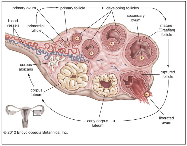
Anatomy of the Ovary
From the regression equation we observe a negative relationship between age and number of eggs produced. In other words, we observe a negative relationship between the Endometrial Thickness and the number of eggs produced.
Declaration of Competing Interest
The research work passed through the research acceptable process which include ethical approval, perused by other researchers etc to this, we assure you that there are no competing interest of any kind.
Acknowledgments
We thank the women that volunteered during this research work, the hospital that hosted the researchers and the community members. Most importantly the health workers that impacted in this study
References
- Amanvermez Ramazan, Tosun Migraci (2016) An update on Ovarian Aging and Ovarian Reserve Test.
- Broekmans FJ, Knauff EAH, Velde ERT, Macklon NS, Fauser BC (2007) Female reproductive ageing: current knowledge and future trends. Trends Endocrinol Metab 18: 58-65.
- Balen A H, Rutherford (2007) Managing anovulatory infertility and polycystic ovary BMJ 335: 663-666.
- Balen AH, Jacobs HS (2003) Infertility in practice. 2nd ed. London: Churchill Livingstone/Harcourt Brace.
- Baskett Thomas F, Calder Andrew A, Arulkumaran Sabaratnam (2014) Munro Kerrs Operative Obsterics E-Book. Elsevier Health Sciences 268.
- Brown SE, Mandelin E, Oehninger S, Toner JP, Seppala M, et (2000) Endometrial glycodelin-A expression in the lutal phase of stimulated ovarian cycles. Fertil Steril 74: 130-133.
- Brown HM, Russell DL (2013) blood and lymphatic vasculature in the ovary: development, function and Hum Reprod Update 20: 29-39.
- Budinetz TH, Benadiva CA, Griffin DW, Engmann LL, Nulsen JC, et (2015) Ovulation rate and cycle characteristics in a subsequent clomiphene citrate cycle after stair-step protocol. Fertil Steril 103: 675-679.
- Chandhry S, Chaudhry K (2020) StaPearls. Statpearls publishing; Treasure Island (FL): Anatomy, Abdomen and Pelvis, Uterus Round Ligament.
- Chandler TM, Machan LS, Cooperberg PL, Harris AC, Chang SD (2009) Mullerian duct anomalies: from diagnosis to intervention. Br J Radiol 82: 1034-1042.
- Colvin Caroline Wingo, Abdullatif Hussein (2013) Anatomy of female puberty: The clinical relevance of developmental changes in the reproductive system. Clinical Anatomy 26: 115-129.
- Daftary Shirish, Chakravarti Sudip (2011) Obsterics, 3rd Edition. Elsevier 1-16.
- Ziegler D, Pirtea P, Galliano D, Cicinelli E, Meldrum D (2016) Optimal uterine anatomy and physiology necessary for normal implantation and placentation steril 105: 844-854.
- Roly ZY, Backhouse B, Cutting A, Tan TY, Sinclair AH, et (2018) The cell biology and molecular genetics of mullerian duct development. Wiley 7: e310.
- Ross M, Pawlina W (2011) Histology: A Text and Atlas (6th Ed.) Lippincott Willams & Wilkins.
- Rzepka-Gorska I, Tarnowski B, Zielinska D, Toloczko- Grabarek A (2006) Premature menopause in patients with BRCA I gene Breast Cancer Res Treat 100: 59-63.
- Siristatidis Charalampos S, Gibreel Ahmed, Basios George, Maheshwari Abha, Bhattacharya Siladitya (2015) Gonadotrophin-releasing hormone agonist protocols for pituitary suppression in assisted reproduction. Cochrane Database of Systematic Reviews Cochrane Database Syst Rev 11.