Hybrid Prisms: Transforming Medical Imaging Technology
© 2024 Friedrich Bjorn Grimm, et al. This is an open-access article distributed under the terms of the Creative Commons Attribution License, which permits unrestricted use, distribution, and reproduction in any medium, provided the original author and source are credited.
Abstract
This research article delves into the groundbreaking use of hybrid prisms in X-ray imaging devices, emphasizing their role in enhancing the precision and efficiency of imaging systems. A hybrid prism, which integrates the functions of a lens and a reflective prism, is a versatile optical component capable of manipulating electromagnetic waves across a spectrum of wavelengths, including visible light and X-rays. This technology significantly improves the focusing and imaging capabilities of X-ray devices, making it indispensable in fields such as medical diagnostics.
Introduction
The evolution of optics from its beginning to the contemporary era has been marked by remarkable milestones and discoveries. From the rudimentary convex lenses of ancient times to the revolutionary insights of Galileo Galilei, Johannes Kepler, and Isaac Newton, the journey of optics has been one of continual refinement and innovation. The advent of X-rays by Conrad Roentgen ushered in a new epoch of imaging possibilities, further propelling the trajectory of optical advancements. Within this dynamic landscape, our research embarks on exploring the transformative potential of hybrid prisms, bridging the realms of lenses and prisms to redefine imaging capabilities across a spectrum of electromagnetic waves.
Description of the Hybrid Prism
The hybrid prism, an innovative optical lens that combines the properties of a lens and a reflective prism, features a rotational body shaped like a rhomboid. This body consists either of a denser glass for visible light or a vacuum for X-rays, surrounded by an optically thinner cladding to facilitate precise object imaging through linked ray paths. While the input and output of light can be refractive or diffractive, the hybrid prism’s two internal totally reflective surfaces cancel out chromatic aberration. For X-rays, with a refractive index less than 1, the vacuum is optically denser than the surrounding material, making refractive lenses typically plano-concave or biconcave. The numerical aperture, representing the lens’s light-gathering ability and resolution, is determined by the ratio of its radius to focal length [1-10].
The invention leverages the combination of a lens and a reflective prism to achieve superior imaging performance across different spectral ranges of electromagnetic waves. A conventional prism refracts light at its boundaries and can cause total internal reflection, changing the light’s direction. In contrast, a lens refracts light rays to converge or diverge, forming images. The hybrid prism surpasses these limitations by utilizing a rotation rhomboid as its core, composed of an optically denser glass for visible light and a vacuum enclosed by glass or metal or for X-ray radiation. This core, with four interfaces to an optically thinner envelope, directs rays along a linked path to form clear images [8].
The rotationally symmetric body of the hybrid prism is designed for X-rays within the wavelength range of 0.1-5 nm. It includes a vacuum interfacing with an optically thinner, dual-part structure: a spindle arranged concentrically and coaxially with the optical axis, and a sleeve positioned radially apart. The spindle’s generating curve comprises straight longitudinal sections with constant inclination angles and, in at least one section, a hyperbola or parabola. The sleeve’s generating curve also consists of straight longitudinal sections with constant inclination angles, incorporating a parabola or ellipse in at least one section. The body’s front and rear boundary surfaces, formed by refractive and/or diffractive correction lenses, deflect X-rays away from the optical axis at the front lens and towards the optical axis at the rear lens, causing total reflection four times at each of the spindle and sleeve boundary surfaces [1,2].
This configuration allows the integration of the condenser and imaging optics of an X-ray microscope into a hybrid collecting prism. It precisely focuses X-rays from a synchrotron with an undulator, forming a brilliant monochromatic parallel beam on a focal point of 0.1 mm or smaller. The hybrid condenser prism acts as an objective for slightly divergent X-rays from a synchrotron, focusing them to a spot of 0.1 mm or less and producing a microscopic image on a CCD sensor [6-8].
The hybrid prism’s design extends beyond optical properties, offering durability, reliability, and ease of integration. Unlike conventional optical components, which are prone to wear and degradation, hybrid prisms withstand extended use in diverse environments. Their robust construction and stable performance make them ideal for applications like space exploration, medical imaging, and industrial inspection. Their compact size and lightweight nature also suit integration into portable devices, broadening their application scope.
Three distinct methodologies are employed to address the issue of low reflectivity in the X-ray domain. These include:
- Grazing Incidence: The introduction of a flatter angle of incidence serves to enhance the reflectivity of surfaces. When the refractive index is less than 1, total reflection can even occur at extremely flat angles of incidence. Consequently, mirrors utilising grazing incidence are frequently deployed in X-ray An illustrative example of an optical apparatus that operates with grazing incidence is the Wolter telescope.
- Multilayer Systems: Mirrors made of multilayer systems are often used for mirrors that have a high reflectivity at steep angles of incidence and are only intended to work at one wavelength. These consist of two different materials that are arranged in alternating layers on top of each other. These multilayer systems are always designed for a specific wavelength and a defined angle of incidence. As a rule, an optically dense and an optically thin medium are used at the corresponding wavelength. The layer thicknesses are matched to each other in such a way that the period duration always corresponds to the wavelength for the intended angle of incidence. Reflection at the optically denser layers leads to constructive One promising coating system, for example, is the combination of silicon and molybdenum for wavelengths around 13.5 nm. Silicon is the optically thin medium and molybdenum the optically denser medium.
- Bragg Reflection: In the case of hard X-rays, the constructive
interference of waves on the crystal lattice described by the Bragg equation can be utilized. This leads to the generation of a diffraction reflection at a crystal at a certain angle and a certain wavelength. However, the intensity of the reflected beam is very low.
Applications of the Hybrid Prism
The study presents a range of applications for the hybrid prism, showcasing its versatility and potential impact in various fields of optics and imaging technology.
These detailed descriptions of the applications demonstrate how the hybrid prism, through converging cavity lenses, can significantly enhance the capabilities of X-ray imaging for various fields.
Focusing Hard X-rays by a Converging Cavity Lens
The longitudinal section below (Figure 1) shows a parallel bundle of hard X-rays that has been homologized by a synchrotron and an undulator to form a parallel bundle with a diameter in the range of 1 to 10 mm. The cavity lens is able to precisely focus a converged X-ray beam onto a focal point (Fd). The parallel X-ray beam, which is represented by the beams (A, B), undergoes a fourfold total reflection before it is precisely focused at the focal point (Fd). The mirror symmetry of the cavity lens enables achromatic imaging by double correction of chromatic aberrations. With hard X-rays, structural geometries smaller than 5 nm are accessible, which opens up new possibilities for microscopy and also for chip production. Critical angles of 0.07 degrees are required for hard X-rays in order to achieve total reflection. If absorption losses are accepted, X-ray imaging is also possible within larger critical angles. The cavity is sealed off from the atmosphere by four optical surfaces (a,b,c,d), whereby a and d can be either refractive or defractive correction lenses in order to homogenize the interlinked beam passage [6,7,11].
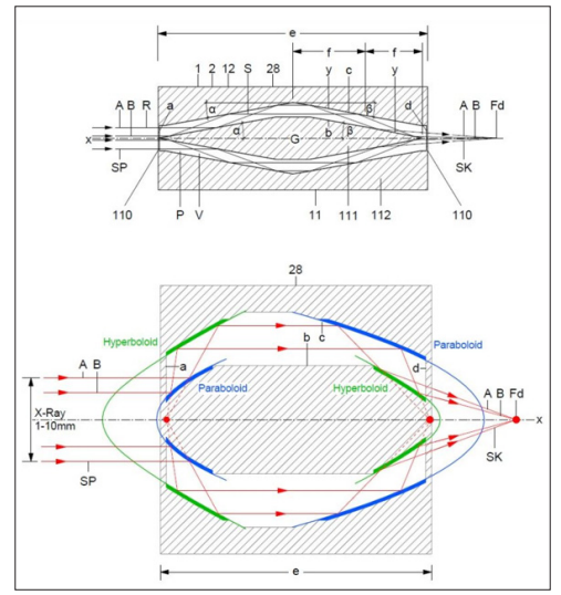
Figure 1: How to focus a diversion bundle of soft x-rays for enlarged X-ray imaging
Soft X-rays Focused by a Converging Cavity Lens
Cavity lenses, capable of focusing radiation across the entire spectrum from infrared to X-rays, form a cavity between an outer rotationally symmetrical body and an inner spindle around an optical axis. For X-rays, the cavity is optically denser than the surrounding matter, giving the refractive index a value just below 1. Soft X-rays, emanating from an X-ray tube as a deflected beam, are focused by the cavity lens. The lens consists of an outer shell and an inner spindle, which is attached to the shell with fastening elements or contactlessly with electromagnets. In the first longitudinal section of the cavity lens, beams A and B are totally reflected at a hyperbolic surface of the spindle, then again at a rotational paraboloid, forming a parallel bundle. This bundle undergoes total reflection at another rotational paraboloid before being focused at the focal point. This interlinked beam path enables a tenfold magnification of the X-ray image on film. Soft X-rays, in combination with a platinum coating, are totally reflected with a critical angle of about 0.25 degree, allowing for a total cavity lens length of only 200 mm in medical X-ray devices [2,6,7,8,11].
Different kinds of X-ray Devices
In medical X-ray devices, the radiation source is formed by an X-ray tube with a point-shaped radiation source which emits a divergent radiation beam with a useful aperture angle of less than or equal to 10 degrees as soft X-rays in the range from 10 keV to 20 keV. Inside the X-ray tube there is an objective for the X-rays, which is designed as a hybrid condenser prism, the front focus of which is arranged congruently with the radiation source of the X-ray tube, which is assumed to be punctiform. The condenser prism is designed to homogenise the X-rays by means of the front correcting lens and to concentrate them at the boundary surfaces of the rotationally symmetric body in an interlinked beam path with quadruple total reflection onto a rear focus of the rear correcting lens. An object formed by a body or part of a body is then X-rayed. The X-ray apparatus can be designed, for example, as a tomograph that rotates around the object so that sharp tomographic images of the object can be obtained on a cylindrical imaging surface by means of a cell detector [1,2,8].
For X-ray microscopes, the optical system has a hybrid collecting prism and is designed to concentrate the monochromatic parallel beam with a beam diameter of 1.0 mm to 10 mm, which is coupled out at a synchrotron with an undulator, as hard X-rays in the range of 10 keV to 125 keV through an objective formed by the hybrid collecting prism onto a focus of the rotationally symmetric body associated with the rear boundary surface. A divergent beam is then projected onto an image surface to obtain a microscopic image of the object irradiated by the parallel beam of X-rays using a CCD sensor of a CCD camera. Alternatively, the optical system of the X-ray microscope can have a hybrid condenser prism. The condenser prism combines the function of a condenser and an imaging objective, whereby the divergent X-ray beam emitted by a synchrotron in the range of 10 keV to 125 keV is first concentrated on a focus of the rotationally symmetric body assigned to the rear boundary surface, in order to subsequently obtain a microscopic image of the object transmitted by the divergent X-ray beam on an image surface by means of the CCD sensor of a CCD camera [1,2,6-9].
Intraocular Prism for Macular Degeneration
An innovative application of the hybrid prism is its use as an intraocular prism for individuals suffering from macular degeneration. Macular degeneration is a prevalent cause of severe visual impairment, affecting a significant portion of the elderly population. The macula, responsible for central vision, may become damaged, leading to a loss of sharp, central vision. Current treatment options have limited success in advanced cases of macular degeneration.
The hybrid intraocular prism addresses this issue by redirecting linked rays within the human eye around the macula. The prism consists of at least two ring-shaped glass bodies arranged concentrically and coaxially with respect to the optical axis. By positioning the focus of the hybrid intraocular prism within the eye at a distance from the retina, a ring-shaped image field
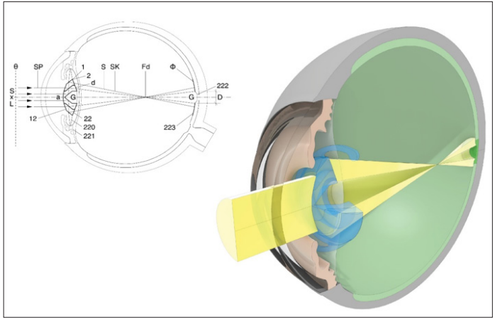
Figure 2
is created around the macula. This allows rays of the linked path to bypass the macula on the inner side of the eye and project a complete image onto the healthy retina surrounding the macula. The hybrid intraocular prism can restore vision and improve the quality of life for individuals with macular degeneration [2,4,5,8].
Future Directions and Challenges Advancements in Hybrid Prism Technology
The future of hybrid prism technology holds exciting opportunities for further innovation and discovery. Continued research and development efforts are expected to provide new insights into the design, fabrication and performance of hybrid prisms, enabling even greater precision and efficiency in light manipulation. Emerging technologies such as metamaterials and photonic crystals may offer new ways to enhance the optical properties of hybrid prisms and open up new applications in areas such as quantum computing and photonics.
Researchers are also exploring novel materials and fabrication techniques for hybrid prisms to achieve even greater precision and efficiency. By harnessing the power of advanced computational algorithms and nanofabrication techniques, scientists can push the boundaries of optical engineering and unlock new possibilities for hybrid prism-based systems. The integration of hybrid prisms with emerging technologies such as artificial intelligence and nanotechnology holds immense potential for expanding their capabilities and applications. By leveraging these synergies, researchers can develop hybrid prism-based systems that are more intelligent, adaptive, and versatile, paving the way for new discoveries and innovations in science and technology.
Addressing Technological Challenges
Despite their numerous advantages, hybrid prisms also face certain technological challenges that must be addressed to realize their full potential. These challenges include manufacturing complexity, cost considerations, and performance optimization in extreme environments. Overcoming these hurdles will require interdisciplinary collaboration and ongoing research efforts to refine fabrication techniques, reduce production costs, and enhance the robustness of hybrid prism-based systems.
While hybrid prisms are designed to minimize chromatic aberrations, focusing X-rays of different wavelengths can still be challenging. If the prism is optimized for a specific wavelength range, X-rays that fall outside this range might not be focused as accurately, potentially causing a small blind spot where the imaging resolution drops.
By addressing these challenges head-on, researchers can unlock new opportunities for advancing optical engineering and harnessing the full potential of hybrid prism technology.
Offering a New Approach to Diagnosis and Treatment State of the Art
The technique of X-ray diagnostics is predicated on the differential absorption of X-rays by the various human tissues. The result of the X-ray examination is an image in digital or analog format, which is then subjected to a medical assessment. Conventional X-ray diagnostics remains the most prevalent method of X-ray diagnostics. It does not provide sectional images; rather, it produces projections in which the radiographed structures are superimposed. Consequently, it is also referred to as “projection radiography.” Computed tomography on the other hand is a relatively recent method that has become an indispensable tool in modern diagnostic imaging, offering a level of detail and precision that is comparable to that of conventional X-ray diagnostics. There exists a wide range of indications for X-ray diagnostics, with the most frequent application being the detection of pathological fractures in hard tissue.
Radiotherapy
Along with surgery and chemotherapy, radiotherapy is one of the most important forms of cancer treatment. Unlike systemic drug- based chemotherapy, which affects the whole body, radiotherapy is a purely local treatment that destroys the tumor only within the radiation field. 3D radiotherapy is a technique in which the radiation field is adapted to the shape and size of the tumor using apertures and filters, so that neighboring tissue structures are protected. 3D radiation is necessary when a tumor is in close proximity to vital organs or structures.
The precision with which 3D-focused X-rays can be directed at individual cancer cells and scattered tumors using a hybrid prism represents a significant advancement in the field of radiotherapy, offering a new dimension of treatment precision and protection of neighboring tissue for 3D irradiation.
Conclusion
The hybrid prism represents a groundbreaking development in optics, offering a versatile and efficient solution for precise imaging of electromagnetic waves in different spectral ranges. Its unique properties, such as the combination of lens and prism functionalities and the ability to focus light and X-rays, make it highly valuable for a wide range of applications. The research article concludes with a discussion on the potential impact of the hybrid prism on the field of optics and its prospects for future research and development [12].
In conclusion, the hybrid prism has the potential to revolutionize optical systems, providing new possibilities for imaging and enhancing various applications. Its ability to focus both light and X-rays opens doors to improved imaging technologies in medicine, research, and various industries. Further research and development in this area are likely to lead to even more exciting advancements and applications for the hybrid prism in the future. As the field of optics continues to evolve, the hybrid prism’s contributions are expected to play a pivotal role in shaping the future of imaging technology and beyond [13].
Reference Signs
|
Hybrid prism |
1 |
Optical system |
2 |
|
Beams |
S |
Optical axis |
x |
|
Example beams |
A,B |
Focus |
Fa-Fd |
|
Divergent beam |
SD |
Radiation source |
Q |
|
Parallel beam |
SP |
Light |
L |
|
Convergent beam |
SK |
X-rays |
R |
|
Rotational rhomboid |
P |
X-ray tube |
21 |
|
Angle of inclination |
α |
Synchrotron |
210 |
|
Tangent angle |
β |
Undulator |
211 |
|
Angle of aperture |
δ |
Object |
Θ |
|
Vitreous body |
10 |
Image area |
Φ |
|
Single element |
100 |
Intraocular prism |
22 |
|
Array |
101 |
Haptic |
220 |
|
Gap |
G |
Capsular bag |
221 |
|
Vacuum |
V |
Macula |
222 |
|
Envelope |
11 |
Retina |
223 |
|
Corrective lens |
110 |
Inner diameter |
D |
|
Spindle |
111 |
Lidar system |
23 |
|
Sleeve |
112 |
Laser |
230 |
|
Anterior interface |
a |
Filter element |
231 |
|
Inner interface |
b,c |
X-ray unit |
25 |
|
Rear interface |
d |
Tomograph |
250 |
|
Length |
e |
Cell detector |
251 |
|
Longitudinal section |
f |
X-ray microscope |
26 |
|
Generating curve |
y |
X-ray telescope |
27 |
|
Fresnel structure |
z |
Objective |
28 |
|
Hybrid collecting prism |
12 |
CCD camera |
280 |
|
Hybrid diverging prism |
13 |
CCD sensor |
281 |
|
Hybrid condenser prism |
14 |
Spot light |
29 |
References
- Grimm F (2021) Hybridprisma als Bauelement für optische Systeme. German Patent No. DE 10 2020 001 448 B3.
- Grimm F (2021) Optical Component Comprising a Hybrid Prism. International Patent No. WO 2021/175910 A1.
- Hong X, Vannoy SJ, Zhang X (2007) Pseudo-accomodative IOL having diffractive zones with varying Alcon Mfg Ltd, International Patent No. WO 2007/092949 A1.
- Lang AJ, Zhao H (2008) Multi-zonal monofocal intraocular lens for correcting optical aberrations. Advanced Medical Optics Inc. US Patent No. US 7 381 221 B2.
- Buralli DA, Federico RJ, Morris GM (2004) Diffractive lenses for vision correction. Apollo Optical Systems Llc https:// google.com/patent/US7025456B2/en.
- Helmholtz-Zentrum Berlin für Materialien und Energie Gmb H, Schneider G (2013) Röntgenmikroskop mit Kondensor- Monochromator-Anordnung hoher spektraler Auflösung. German Patent No. DE 10 2005 056 404 B4.
- Wang Y, Yun W (2004) Phase contrast microscope for short wavelength radiation and imaging X radia Inc, US Patent No. US 2004/0125442 A1.
- Niemann Bastian (1997) Condenser monochromator arrangement for X-rays. German Patent DE 197 00 615 A1.
- Grimm F (2020) Kameramoduleinheit für German Patent No. DE 10 2017 011 352 B4.
- Physikalische Blätter 57 (2001) Nr 1:
- Pareschi G, Spiga D, Pelliciari C (2021) X-ray Telescopes Based on Wolter-I Optics 12.
- Andrew J Morgan, Mauro Prasciolu, Andrzej Andrejczuk, Jacek Krzywinski, Alke Meents, et (2015) High numerical aperture multilayer Laue lenses.
- Frank Seiboth, Andreas Schropp, Maria Scholz, Felix Wittwer, Christian Rödel, et (2017) Perfect X-ray focusing via fitting corrective glasses to aberrated optics. Nat Commun 8: 14623.