Cardiac Auscultation: A Practical Method for Instruction and Learning
© 2024 Morton E Tavel, et al. This is an open-access article distributed under the terms of the Creative Commons Attribution License, which permits unrestricted use, distribution, and reproduction in any medium, provided the original author and source are credited.
Abstract
In response to the worldwide deficiency in the use of standard acoustic stethoscopes for the analysis of cardiac disorders, we have developed a cost-free platform for learning and teaching of cardiac auscultation by combining graphic images with actual patient recordings.
Recent studies have cited the obvious fact that the talent for the use of standard stethoscopes in analysis of cardiac disorders has steadily declined, but proficiency in this technique remains important. As indicated recently by Azmeen et al and Voin et al. The high prevalence of cardiac diseases around the world continues to provide a need for quick, easy and cost-effective approaches to diagnose heart disease [1,2]. Auscultation and interpretation of heart sounds using the stethoscope is inexpensive, should require minimal to advanced training, and is widely available to all healthcare providers working in urban environments or, especially, in medically underserved rural areas. Unfortunately, however, many physicians and health care providers are uncomfortable and often inaccurate in their ability to identify normal and abnormal heart sounds.
Why is This?
Many years ago, upon my introduction to cardiac auscultation, it was the usual custom for our mentors to instruct us by applying the stethoscope to a specific point on the chest of a cardiac patient. The instructor would then inform us of a specific sound that he had just heard. I may or may not have heard the alleged sound, but often to please the instructor, I and others would often blindly “affirm” that we had heard the said sound phenomenon. This admittedly uncertain process was duplicated repeatedly with most of our fellow trainees. This method of learning has persisted for many years, and it has provided at least one reason why most clinicians are unable to properly employ and instruct though the use of the stethoscope. Adding to this uncertainty, even our mentors may have incorrectly identified or missed some of the sounds that may- or may not- have been present.
Long ago, after struggling with this laborious process of learning how to use the stethoscope in this way, I acquired this ability primarily through a method of self-teaching with visual displays (phonocardiograms), combined with near simultaneous listening with the stethoscope. Although phonocardiography is a very useful teaching and diagnostic tool, and it had been used extensively to form a basis for modern auscultation, it is a rather laborious technique that is not easily and generally adaptable, and for the past several years it has been largely abandoned and neglected as a teaching tool [3]. Thus, in the absence of this method for accurate hearing and interpretation of sounds, clinicians now usually abandon the stethoscope, instead relying on other- but expensive- sonar and doppler techniques. Moreover, a simple stethoscope may provide even more information that is unavailable through these former methods. For example, the presence of audible third or fourth heart sounds can provide more information than that of imaging or doppler studies. In addition, the presence and intensity of many cardiac murmurs may be more clinically relevant than the presence of a small jet of mitral or aortic regurgitation as shown by doppler techniques. The judicious use of the stethoscope alone can allow identification of innocent murmurs and, in the process, can avoid the need to resort to complex testing.
To circumvent this problem of suboptimal cardiac auscultation, we have developed a technique that employs actual sound recordings from patients that demonstrate a wide variety of normal and abnormal cardiovascular sounds. Like phonocardiography, this method displays graphically recorded sounds, but by contrast, this is combined with a moving cursor that tracks and plays the sounds as they occur (Figure 1). It enables the trainee to identify the sounds visually that, provides a learning experience that resembles the earlier method that coupled sounds with graphically displayed images. This system was derived from actual patients with various cardiovascular maladies, and when used with ear buds or standard headsets, the sound quality closely simulates that heard from conventional stethoscopes. After one hears and identifies the sounds as presented, it enables the creation of a corresponding mental image to use when one later encounters actual patients. To provide maximum availability, we have presented this platform though a free link to the following internet location: https:// www.screencast.com/users/Auscultation/folders/Default/media/ c4fde766-589c-4891-b82c-43ad93e06b50 This technique is also applicable to both teachers and students, and, although readily available for individual use, given proper equipment, it can even be made available to multiple simultaneous listeners.
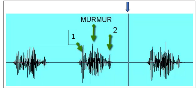
Figure 1: A Typical Graphic Display Showing Labeled Sound Components. The Moving Cursor (Vertical Line) Sweeps Across Simultaneously with the Audible Events as they Occur. This Recording was Made from a Male with Severe Valvular Aortic Stenosis
References
- Azmeen A, Vakilzadian H, Haider H, Mathers DH, Zimmerman R (2023) Heart sounds: Past, present, and future from a technological and clinical perspective - a systematic review. Proc Inst Mech Eng 237: 669-682.
- Voin V, Oskonuian RJ, Louka M, Tubbs RS (2017) Auscultation of the heart: The Basics with Anatomical Correlation. Clin Anat 30: 58-60.
- Tavel ME (1985) Clinical Phonocardiography and External Pulse Recording. (4th Ed), Year Book Medical Publishers,