A Green Approach on Structural, Optical and Anti-Bacterial and Anti- Fungal Studies of Gold Nanoparticle with Fenugreek Seeds
© 2023 Deepa Rosh Tom, et al. This is an open-access article distributed under the terms of the Creative Commons Attribution License, which permits unrestricted use, distribution, and reproduction in any medium, provided the original author and source are credited.
Abstract
Recently we are more conscious with covid-19 virus and other pathogens too. In order to prevent us from these micro-organisms we are using face mask, hand gloves etc which are coated with nano materials. Nano particles having antibacterial effect coated on materials will help to protect against pathogens. In the current research gold nano particles have the capability to fight against micro-organisms which is synthesized by very simple method called green synthesis. Nano particles obtained by green method is user-friendly as it is less toxic and some are non-toxic.
The resultant nano particle is of purple in colour with average crystalline size of 51nm which was found by XRD and the sharp peak says that it is FCC crystalline structure. The SEM, FTIR, FT-RAMAN, UV, PL analysis were done and the material showed high anti-Bacterial and Antifungal Activity.
Introduction
Bioreduction is one of the most easy and eco-approach for the creation of gold nanoparticles. Even though nano sized particles can be made by physical or chemical synthesis, Green synthesis is the unaffected way to the mankind. Nano-sized particles can be seen in everyone?s day-to-day life. For a person from teaching background, he is always playing with nano sized chalk powder. If he is a mason then, always be with cement which consists of nano size one. So we can see the nano- things everywhere and from ancient time onwards we are known to it. But all nano sized materials cannot be called as nanoparticles unless its range is in between 1nm to 100 nm. It is possible to heal the genetic issues by fixing the damage genes. Nanotechnology could be used for making drugs at a molecular level which will become more effective. Thus reduces the side effect? [1].
?The topic there is plenty of room at the bottom was one of the famous talk of Richard Feynman which was the ground of Nanotechnology and Nanoscience in 1959. In his talk, he predicted that one day the encyclopedia would fit on the head of pin and a library having all the books in the world wide would fit in the three square yards. A technology is called as nanotechnology only if it have the ability to manipulate, create and use the structures, devices on the atomic scale.?[2]. The authors studied the plant-mediated synthesis of silver and gold nanoparticles and their applications [3].
Plant parts such as leaf, root, latex, seed, and stem are being used for metal nanoparticles synthesis. [3] Gold nanoparticles are the most attractive member of metallic nanoparticles due to their huge applications in fields such as catalysis [4], drug delivery [5], imaging, bio-sensing, gene expression, and disease diagnosis [6- 10]. The presence of biomolecules in fenugreek seeds will act as reducing agent and convert gold metal ions into gold nano atoms by green synthesis. Biosynthesis of Controllable Size and Shape Gold Nanoparticles by Black Seed (Nigella Sativa) Extract have been presented [7]. Green synthesis of gold nanoparticles using Citrus fruits (Citrus limon, Citrus reticulata and Citrus sinensis) aqueous extract and Fenugreek Seed extract have also been looked into [10,11].
Synthesis of Gold Nanoparticles
Powdered fenugreek (10g) is measured by using electronic balance and added to 150 ml of distilled water. Stir it for 1 hour and allow to filter the solution twice by using a filter paper. 1 mole of gold chloride is added into 50 ml of distilled water in another beaker. Mix both the solutions in the beakers and allow for 2 hours of stirring. The solution is then centrifuged for 6000 rpm, to separate the solid from the solvent. The material was washed several times with distilled water and later washed with ethanol. The formation of gold nanoparticles is indicated by a shift in the color of the solution from pale brown to violet.
The bio components contain in the fenugreek are responsible for the immediate reduction of ions and formation of gold nanoparticles. The violet coloured material is then left for heating at 180 °C for 8 hours. The dried material obtained was grinded to make it a fine powder and calcinated at 450 °C. The final product prepared is then given out for characterization studies like XRD, UV-VIS-NIR, FTIR, SEM/EDX and PL and FT-Raman.
Result and Discussion
In figure (1b) the intense diffraction peaks were observed at 2? values of 38.1194°, 44.3255°, 64.4876°, and 77.4877°, which were indexed to the (111), (200), (220) and (311) reflections of polycrystalline face-centered cubic (FCC) structure of metallic gold respectively. These values are matching with Joint Committee on Powder Diffraction Standards (JCPDS no. 04- 0784) and saying that synthesized AuNPs are of pure crystalline gold with average crystalline size of 51nm. The XRD pattern is of sharp peak thus clearly shows that the AuNPs were crystalline in nature. The surface morphology and size of the Au NPs was analysed by Scanning Electron Microscope. SEM image had shown individual Au NPs as well as number of aggregates. From the images fig (1c) it is evident that the morphology of Au NP is nearly roughly spherical shaped in clusters and shows the size of the Au NPs ranging from 40-80 nm. The energy dispersive X-Ray Spectrum of the product is shown in the figure (1d). The atomic percentages obtained from the spectrum of the Au and O are 66.93 and 33.07 % respectively. The absence of any other peaks suggests that the purity of the product is reasonably good [11].
The method for determination of Gold NPs formation and their stability is carried out in the wavelength range of 300-800 nm at different time intervals. Spectrophotometric absorptions in the wavelength ranges of 500-560 nm are used in characterizing the gold nanoparticles. In the present studies of UV fig (2b), the absorption peak is obtained at 550nm which is due to the surface plasmon resonance of the smallest particle size of the material. During the synthesis the colloidal solution showed intense colour due to surface plasmon resonance arising from the collective oscillation of the free conduction electrons induced by an interacting electromagnetic field. The position of SPR band is sensitive to the particle size, shape, refractive index and its interaction with medium [11].
Au NPs exhibit the local electric field caused by the localized surface plasmon resonance behaviour [13]. Figure (2a) shows the PL spectrum of gold nanoparticles with fenugreek. The PL spectrum was characterized by the presence of two peaks. First peak is sharp showing the absorption with the peak centered at 370 nm. The second peak resulted the emission at 550 nm showing broad and intense peak due to the functionalization of Au nanoparticles with bioactive in fenugreek. It has been known that the intensity of the plasmon peak is greatly dependent on the size and shape of Au NPs [12]. As the gold crystallite size becomes smaller, the PL intensity becomes higher and stronger [13]. FTIR is a structural method which measures infrared intensity v/s wavelength or wave number of light. It was used to analyse the bio molecule and also bonding interaction between molecules of the gold nanoparticles.
FTIR measurements were carried out to identify the potential functional groups of the biomolecules in the fenugreek extract responsible for the reduction of the gold metal ions to gold nanoparticles. FTIR spectra of the green synthesized fenugreek gold nano particles are shown in the figure (2c). The FTIR spectrum of the bio-reduced gold nanoparticles had the adsorption peaks located at about 3440, 2370, 1715, 1641, 1086cm-1.
The band at 3440 cm-1 corresponds to stretching vibration of O-H group. The IR band due to C=O stretch is observed at 1715 cm1. The very strong absorption band at 1,641 cm-1 is identified as the amide I and arises due to the carbonyl stretching vibrations in the amide linkages of the proteins. The presence of bands at 1641 and 1086 cm-1 indicates that gold NPs are possibly bound to proteins through carboxylate group. The C-OH vibration of proteins is observed at 1086cm1. Raman spectroscopy is an important method which reveals the molecular level information on composition and structure of cellular components. The characteristic peak positions of the gold nanoparticles were determined in the raman region of interest. There are well defined peaks in the spectrum figure (2d) which can be assigned to different bonds. Raman spectrum convey a band at 570 cm- 1which indicates the N-C=O group and found in amides. Similarly another band at 440cm-1 assigned to skeletal deformation of C-N-C and the band 340 cm-1 indicates C-Cl bond.
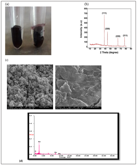
Figure 1: (a) Gold Nanoparticle (b) XRD graph (c) SEM image(d) Energy Dispersive Absorption X-ray Analysis
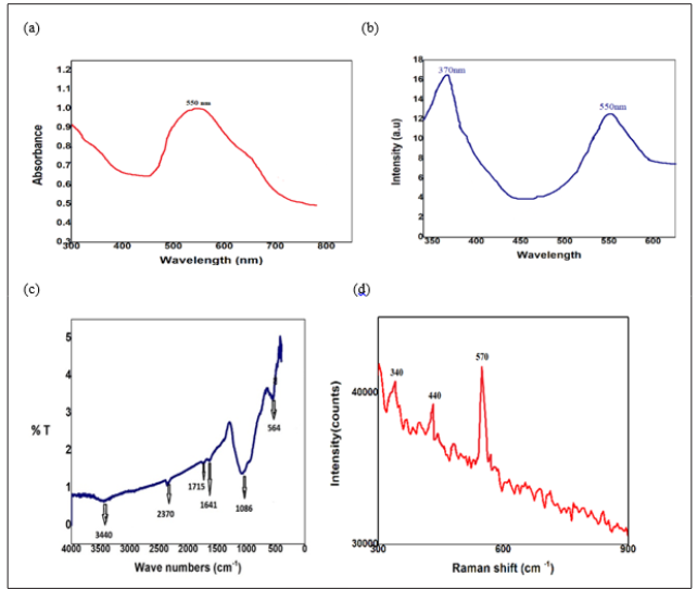
Figure 2: (a) Photoluminescence (b) Spectral UV - VIS - NIR graph (c) Fourier Transform-Infrared Spectroscopy (d) Fourier
Transform-Raman Spectroscopy
Anti -Bacterial Studies
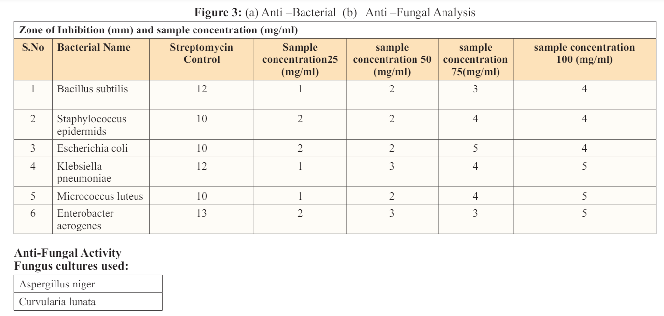
In the current project, gold nanoparticles 40 to 80 nm in size exhibited high antibacterial activity. The result disclose that it was influential against Gram-positive bacteria and Gram-negative bacteria. But As the concentration of the sample increases from 25mg/ ml to 100mg/ml, it is noted that the zone of inhibition around the well is also increasing. The antibacterial mechanism demonstrated that gold NPs can be the next therapy against this enteric bacterium.
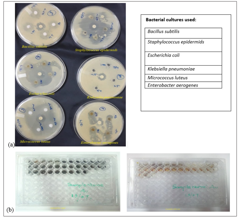
Preparation of fungal spore
Broth micro dilution method consists of four steps for the study of antifungal assay. In this study, Fluconazole was taken as positive control and MIC was calculated.
1.Curvularia lunata, Aspergillus niger were grown on SDA slants at 28°C for 10 days and stored in refrigerator for future use.
2.Gold nanoparticle were dissolved in water and added 2% of DMSO.
3.Each well was inoculated with 5 μl of suspension containing approximately 104spore/ml of fungi.
4.The plate was kept in incubator at 37°C for 20 hours.
5.The plate was taken outside of the incubator and checked for fungal growth. The lowest extract concentration showed no visible fungal growth after incubation time.
The following test fungal strains were used for the experiment: Curvularia lunata 46/01, Aspergillus niger MTCC 1344. About two fungal pathogens which could cause disease in both animals and plants were considered for the present study. Fluconazole was used as a positive control. In the case of Au MIC value was significantly found in Curvularia lunata 46/01 (21.25 μg/ml) and Aspergillus niger (25.25 μg/ml) [14-20].
Conclusion
The purpose of this study was to evaluate the potential of fenugreek extract to synthesize AuNPs through green chemistry. The biosynthesized AuNPs were characterized by different techniques such as UV-Visible, XRD, FT-IR and SEM, PL and FT-Raman. The biological activities of synthesized GNPs such as anti-bacterial activities were evaluated by standard method.
In this study, biological GNPs were successfully synthesized from the extract of fenugreek by the simple, cost-effective and eco-friendly approach. These biosynthesized GNPs were mostly spherical with a size range of 3-80 nm, and their crystallites confirmed by XRD pattern. FT-IR analysis confirmed the presence of phytocompounds involved in reduction and stabilization of nanoparticles. The biogenic GNPs displayed remarkable cytotoxic properties and significant antibacterial activity.
Declaration of Competing Interest
The authors declare that they have no known competing financial interests or personal relationships that could have appeared to influence the work reported in this paper.
Acknowledgement
The research facility provided by the PPSU University is so gratefully acknowledged.
References
1. Chris Binns (2010) Introduction to Nanoscience and Nanotechnology. Wiley 1-416.
2. M A Shah, Tokeer Ahmed (2010) Principles of Nanoscience and Nanotechnology. Narosa publishing house 1-220.
3. SK Yadav, V Kumar (2009) Plant-mediated synthesis of silver and gold nanoparticles and their applications. Journal of Chemical Technology and Biotechnology 84: 151-157.
4. D Astruc, M C Daniel (2004) Gold Nanoparticles: Assembly, Supramolecular Chemistry, Quantum Size-Related Properties and Applications toward Biology, Catalysis and Nanotechnology. Chemical Reviews 104: 293-346.
5. Ghosh P, Gang Han, Mrinmoy De, Chae Kyu Kim, Vincent M Rotello (2008) Gold nanoparticles in delivery applications. Advanced Drug Delivery Reviews 60: 1307- 1315.
6. Anil Kumar, Bhargavi M B, Xing-Jie Liang (2011) Gold Nanoparticles: Promising Nanomaterials for the Diagnosis of Cancer and HIV/AIDS. Journal of Nanomaterials, 2011: 17.
7. Ahmed Fragoon, Jianjun Li, Jian Zhu, Junwu Zhao (2012) Biosynthesis of Controllable Size and Shape Gold Nanoparticles by Black Seed (Nigella Sativa) Extract. Nanoscience and Nanotechnology 12: 1-9.
8. Farrukh DMA (2012) Atomic Absorption Spectroscopy. In Tech 1-272.
9. F Zahir, S Honary (2013) Effect of Zeta Potential on the Properties of Nano-Drug Delivery Systems - A Review (Part 2). Tropical Journal of Pharmaceutical Research 12: 265-273.
10. Sujitha MV, S Kannan (2013) Green synthesis of gold nanoparticles using Citrus fruits (Citrus limon, Citrus reticulata and Citrus sinensis) aqueous extract and its characterization. Spectrochimica Acta Part A: Molecular and Biomolecular Spectroscopy 102: 15-23.
11. Ahmed Fragoon, Amal Mamoun, lamiaa Frah, Shahinaz Abd-Alwahab (2016) Biosynthesis of Gold Nanoparticle by Fenugreek Seed (trigonella foenum) Extract. ICCNEEE 1: 50-55.
12. Chan K, Goh BT, Rahman SA, Muhamad MR, Dee CF, et al. (2012) Annealing effect on the structural and optical properties of embedded Au nanoparticles in silicon suboxide films. Vacuum 86: 1367-1372.
13. Tengku Sarah Tengku Amran, Md Roslan Hashim, Nihad K Ali Al-Obaidi, Hanani Yazid Rohana Adnan (2013) Optical absorption and photoluminescence studies of gold nanoparticles deposited on porous silicon. Nanoscale Research Letters 8: 35
14. Madar Z, Rachel A, Shlomith S, Joseph A (1988) Glucose lowering effect of fenugreek in non-insulin dependent diabetics. Eur J Clin Nutr 42: 51-54.
15. Sharma RD, Raghuram TC, Rao NS (1990) Effect of fenugreek seeds on blood glucose and serum lipids in type I diabetes. Eur J Clin Nutr 44: 301-306.
16. Thermo Nicolet (2001) Introduction to Fourier Transform Infrared Spectroscopy. Thermo Nicolet corporation https://www.scirp.org/(S(czeh2tfqw2orz553k1w0r45))/ reference/referencespapers.aspx?referenceid=2512118.
17. Ruquan Ye, Andrew R Barren (2011) Photoluminescence Spectroscopy and its Applications. openStax- CNX module m38357: 1-11.
18. Daniel M C, Astruc D (2004) Gold nanoparticles: assembly, supramolecular chemistry, quantum-size-related properties, and applications toward biology, catalysis, and nanotechnology. Chem Rev 104: 293-346.
19. Fenugreek production in India https://en.wikipedia.org/wiki/Fenugreek_production_in_ India.
20. Spice: Fenugreek seeds. Spices & Medicinal Herbs
http://www.spicesmedicinalherbs.com/fenugreek-seeds- trigonella-foenum-graecum.html.