Author(s): Omar Lazrek*, Moncef Boufettal, Rida Allah Bassir, Moulay Omar Lamrani, Mohammed Kharmaz and Mohamed Salah Berrada
Tuberculous tenosynovitis is a rare localization of tuberculosis. The diagnosis is often late due to often poor and chronic clinical manifestations. We report a case of tuberculous tenosynovitis of the 4th finger of the right hand treated surgically at the department of traumatology and orthopedic surgery of the Ibn Sina university hospital in Rabat.
Rice bodies are described as fibrin bodies typically found in patients with inflammatory joint disease, tuberculous arthritis, and tuberculous tenosynovitis. Its unusual presentations such as tuberculous tenosynovitis are often unrecognized and are associated with diagnostic and therapeutic delay, especially since formal histological proof can sometimes be lacking. We report the case of a 44-year-old woman presenting a mass of the palmar face of the base of the 4th finger. The patient was treated in the Department of Trauma and Orthopedic Surgery at Ibn Sina University Hospital in Rabat. This case represents the only case described in the literature.
44-year-old patient, housewife, followed for iron deficiency anemia under treatment, was referred by her attending physician for painful swelling of my palmar face of the 4th finger of the right hand opposite P2, evolving for 1 year, with extensions at the base of the 4th finger the whole evolving in a feverish context and conservation of the general condition. There was no notion of trauma. The physical examination objectified the soft mass of the 4th finger measuring 1 x 4 cm, with limitation of flexion at 90 ° and sign of inflammation opposite suggesting tenosynovitis of the flexor of the right 4th finger. Biology revealed an inflammatory syndrome with ESR at 50 and hyperleukocytosis at 10,000. The intradermal reaction to tuberculin and the chest roentgenogram were normal. Radiography of the right hand showed thickening of the soft tissue of the right 4th finger without apparent calcification . A joint ultrasound showed a heterogeneous hypoechoic lesion, faintly taking color on Doppler, encompassing the superficial and deep flexor tendons, extended to the palmar face facing the 4th ray suggesting a Tenosynovial giant cell tumor of the sheath of the flexor tendons of the 4th finger. By default of means the MRI could not be performed. The indication for surgical exploration was then asked. The patient was placed in the supine position with the upper right limb on a support. Then, a surgical exploration was performed under locoregional anesthesia. A Z incision was made from the distal fold of the hand to the crease of the proximal interphalangeal joint (figure 1), the opening of the flexor sheath found the presence of several grains of rice which were removed (figure 2). . a complete excision of the sheath was performed while respecting the neurovascular structures. Pathological examination found a synovial mucosa harboring granulomas of varying size made up of epithelioid cells and giant cells without caseous necrosis, first suggesting a tuberculous origin (figure3). Culture and PCR confirmed the tuberculosis origin of the hand. Thus, the patient was treated with isoniazid (INH), rifampicin (RIF), pyrazinamide (PZA) and ethambutol (EMB) for two months then with INH and RIF for four months. The postoperative follow-up was simple, without notable complications. The evolution was marked by a complete recovery of the flexion of the 4th finger without noticeable recurrence.

Figure 1: Z-cut incision from the distal hand crease to the crease of the proximal interphalangeal joint
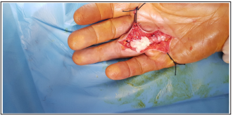
Figure 2: macroscopic appearance of rice grains

Figure 3: Anatomopathological aspect of rice grains
Several authors have speculated on the nature of rice bodies. Albrecht et al. reported that the fibrous bodies of rice represent an end product of synovial inflammation, proliferation and subsequent secondary degeneration [1]. Cheung et al. suggested that the rice bodies originated from infarcted synovial cells, and that these cells spread into joint fluid or bursitis [2]. Berg et al. suggested, in an electron microscopic study of rice bodies obtained from joints of patients with rheumatoid arthritis, that nonvascularized rice bodies may have formed from novo as part of an inflammatory reaction of the synovial fluid [3]. In our case, the cause of the formation of a rice body was of tuberculous origin.
The involvement of the musculoskeletal system represents 1 to 5% of extra-pulmonary manifestations of tuberculosis. Tuberculous tenosynovitis presents 5% of osteoarticular tuberculosis and it predominates in the wrist and on the palmar face of the hand [4- 5]. The flexors are still much more frequent than the extensors. The clinician rarely mentions the diagnosis of tuberculosis when faced with a picture of chronic tenosynovitis and the diagnostic delay is often long. Our patient did not consult until 1 year after the onset of the disease, which testifies to the insidious evolution of the symptoms.
Tuberculosis infection primarily affects the respiratory system. The infection spreads by the lymphohematogenous route and extra-pulmonary involvement occurs at a rate of approximately 14%. The most common place in the musculoskeletal system is the vertebra with an occurrence rate of 30-50%. It can also affect the pelvis, ankle, and wrist. Involvement of the tendon sheath is rare. it is generally due to contamination by the hematogenous route. Lymphogenic or direct contamination can also lead to infection [6] In our patient, tenosynovitis was primary since no other infection, neither contiguous nor distant, was identified.
Biological blood tests have little diagnostic value. An increase in the sedimentation rate (VS) of between 25 and 100 mm 1 / h is present in the vast majority of cases. Nevertheless, SV is normal in 10% to 20% of cases. The skin reaction is generally positive but does not exclude the diagnosis in case of negativity [7]. Ultrasound is a useful examination to confirm the diagnosis of tenosynovitis and show its extension as in the case of our patient. It makes it possible to objectify an increase in the volume of the synovial sheath, forming a sleeve around the tendon. Tendon thickening or abscessing fluid collection may be observed [8]. The MRI is certainly the most useful and the most sensitive examination, the granulomatous synovium of the tendon sheaths is typically of intermediate signal in T1, enhanced by the injection of gadolinium and in hyper signal in T2. The effusion is in hypo signal T1 and hyper signal T2. MRI shows synovial proliferation but also, in some cases, abscess formation and destruction of adjacent bones [9]. Bacteriological evidence is only present during direct examination in 20% of cases and cultures are negative in 35 to 45% of cases [10]. Gene amplification methods from synovial fluid are more sensitive and allow rapid and specific detection of BK [4]. The absence of BK in cultures cannot and should not invalidate the diagnosis.
The treatment is based on an initial quadruple therapy combining INH, RIF, PZA and EMB for 2 months. After 2 months and in the absence of resistance, the treatment becomes a dual therapy combining INH and RIF. The minimum duration of antituberculosis treatment in osteoarticular tuberculosis remains poorly codified at present. The various studies of the literature and international recommendations in particular those of the Center for Disease Control and Prevention and those of the Superior Council of Public Hygiene of France indicate that the 6-month treatment is as effective as a long-term treatment. duration [11].
Current recommendations for the treatment of bone tuberculosis include an initial phase of isoniazid, rifampicin, pyrazinamide and ethambutol lasting 2 months, followed by a regimen of isoniazid and rifampicin of 6 to 9 months. [12]. In our patient, the duration of treatment was only 6 months but the evolution was very favorable with a follow-up of one year.
Tuberculosis infections of the hand, although rare, are still regularly encountered. The course is insidious and the diagnosis remains difficult at the onset stage. Laboratory examinations, ultrasound and MRI can guide the diagnosis. The definitive diagnosis is provided by the histological study. Treatment is based on antituberculosis chemotherapy, surgical treatment has become much rarer, to be discussed on a case-by-case basis.
Conflicts of interest
The authors declare no conflict of interest.
Contributions from the authors
All the authors contributed to the realization of this research work. All authors have read and approved the final version of the manuscript.