Author(s): Daniel Benharroch
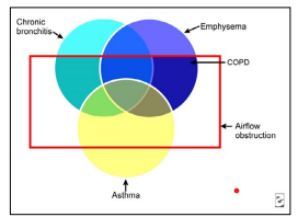
Clinically grouped together
Overlapping features of damage
One common extrinsic trigger:
cigarette smoking
Environmental pollutants increase the incidence of morbidity
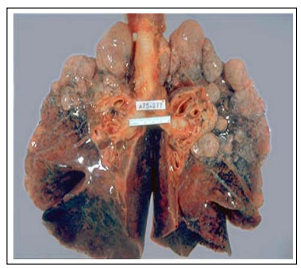
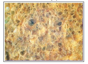
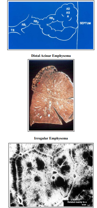
Abnormal enlargement of the alveoli and/or respiratory
bronchioles.
Destruction of alveolar septa.
Absence of significant fibrosis
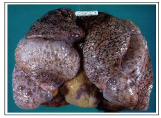
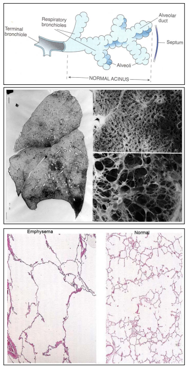

Upper lobes (apical segments) are mainly damaged, in frequency and severity.
Mainly in heavy smokers.
Often together with chronic bronchitis.
In severe cases, if includes distal aspect of the acinus: panacinar emphysema.
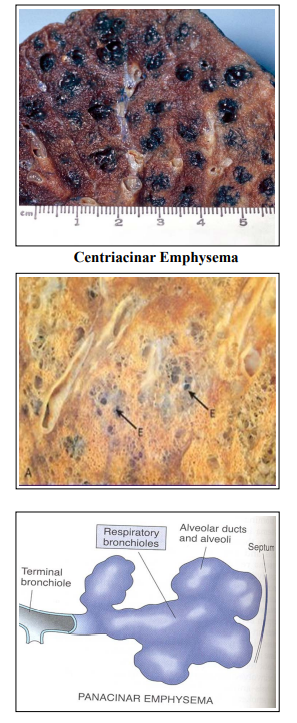
The prefix “pan” concerns the whole acinus. Involves mainly the anterior-lower aspect of the lung. Type associated with alpha1-antitrypsin defi-ciency.
• Compensatory hyperinflation “emphysema”
• Obstructive overinflation “emphysema”
• Bullous “emphysema” (>1cm)
• Interstitial “emphysema”
• Senile “emphysema”
• The definition is clinical: productive cough for two
consecutive years -
• At least three months each year.
• In advanced stages (heavy smokers): chronic airway
obstruction and emphysema.
• Chronic irritation by inhaled substances (90% are smokers).
• Bacterial and viral infection - triggering acute exacerbation.
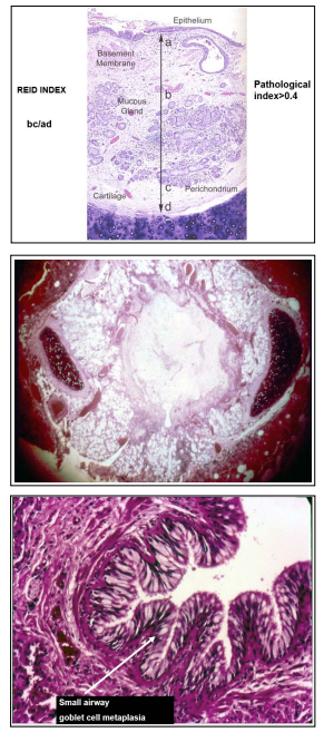
Microscopical features:
Major increase in size of mucous glands
Measured by Reid index
Microscopical features:
Goblet cell metaplasia
Mucous plugging
Inflammatory infiltrate
Fibrosis (bronchiolitis obliterans)

Variable bronchoconstriction and airflow limitation that is at least partly reversible, either spontaneously or with treatment
• Atopic asthma
- Genetic predisposition to type I hypersensitivity.
• Non-atopic asthma
- Virus induced inflammation, lowering the threshold of
the sub-epithelial vagal receptors to irritants
• Drug induced asthma.
• Occupational asthma

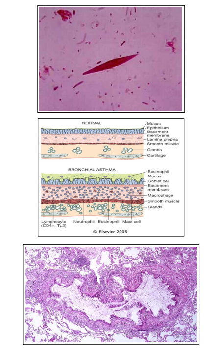
(morphological changes)
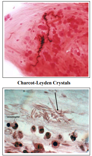
• Tenacious mucous plugs containing:
Curschmann spirals (whorls of shed epithelium)
Numerous eosinophils
Charcot-Leyden crystals (eosinophil membrane protein)
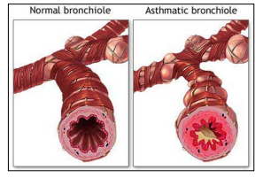
• “Airway remodeling”:
Thickening of the basement membrane of the
bronchial epithelium
Edema and inflammation in bronchial wall Hypertrophy
of bronchial wall muscle