Author(s): <p>Amir Shafiruddin Nordin, Wan Mohd Azizi Wan Sulaiman, Nur Athirah Othman Basri, Rushduddin Al Jufri Roosli, Muhamad Zakuan Abdullah, Jamaliah Mohd Isa, Norfarah Ashikin Baharuddin Anthony and Henkie Isahwan Ahmad Mulyadi Lai*</p>
Hirudinaria manillensis, a freshwater leech endemic to Peninsular Malaysia, exhibits promising antimicrobial and biochemical properties. Its saliva serves as a model for studying annelid biology in leech therapy. This study aimed to assess the protein concentration and antimicrobial activity of crude leech saliva extracts (CLSE) under varying starvation conditions. CLSE was collected by feeding leeches a phagostimulatory solution through a parafilm membrane. The total protein concentration was measured using the Bradford assay, and antimicrobial activity was evaluated through disc diffusion and minimal inhibitory concentration tests. Our findings indicate that protein production and CLSE secretion are influenced by starvation duration. Protein concentration in CLSE significantly decreased from 49.8 mg/L ± 0.033 at 2 weeks to 40.34 mg/L ± 0.053 at 4 weeks, with a drastic reduction to 24.45 mg/L ± 0.003 at 8 weeks, likely due to physiological exhaustion. Microbial activity tests revealed that CLSE collected after a 4 week starvation period exhibited significant inhibition against gram negative strain: Escherichia coli, Klebsiella pneumoniae, and Salmonella enterica, but not Staphylococcus aureus. Beyond the fourth week, no significant changes in inhibitory zones were observed. These results demonstrate that the starvation period significantly affects both protein concentration and antimicrobial efficacy of CLSE. Further investigation into the relationship between starvation duration and protein concentration in relation to microbial activity is necessary. Obtaining sufficient CLSE volumes for comprehensive analysis remains a challenge.
Leeches are widely used as a model organism in toxicology, physiology, neurobiology, biochemistry, histology and many other fields of study. The history of clinical and medical use of leeches can be traced back to the 5th century BC, with significant usage in the 19th, followed by a decline in the early 20th century [1].
The Asian buffalo leech, Hirudinaria manillensis also known as the Asian medicinal leech, is commonly found in natural water bodies of the Southeast Asian region. Adults reach a maximum width of 13 mm and length of 130 - 140 mm, and can stretch up to 200 mm when fully extended [2]. They inhabit the Indo-West Pacific region in slow moving or stagnant waters, typically found in rice paddies, swamps, and sluggish streams. In cattle-rearing countries within the region, they are very frequently found attached to the bellies of livestock, feeding on their blood [3].
In Malaysia, freshwater leeches with Hirudinaria manillensis-like morphology are commonly cultured in farms for use in traditional medicine, leech therapy for blood-related problems and as sources of medicinally bioactive ingredients. Molecular classification places Hirudinaria as a sister taxon to Goddardobdella [4-10].
Hirudinaria manillensis displays various fascinating behavioural and physiological characteristics that are of interest to evolutionary, biochemical and pharmaceutical studies. Unline oligochaetes, which typically have 2 suckers at each end of the body, Hirudinaria manillensis has independent internal and external body segmentations and a robust muscle coelom, making it an excellent model for studying the evolution of the annelid body plan [11].
According to Wagner R, the leech’s ability to overcome blood clotting while feeding and to maintain the blood in a liquid state for extended periods in their crops had attracted the attention of traditional therapists, physicians and researchers as an effective remedy for managing coagulation disorders. Leeches have been the subject of many scientific researches aimed at identifying and characterizing the blood-affecting constituents, especially peptides and proteins, in their salivary gland secretions [12].
This preliminary study aimed to investigate the effectiveness of the starvation period on the total protein concentration and microbial activity using crude leech saliva extract (CLSE) from salivary gland of Hirudinaria manillensis.
A total of 30 leeches used in the study were purchased from suppliers from around Kuala Lumpur and Selangor. The leech used in this study is a type of buffalo leech from Hirudinaria manillensis species. These leeches are easily found in swamp areas and around rice fields found throughout Peninsular Malaysia. Leeches were then stored in small glass jars filled with hill water taken from the area around Hulu Langat. Water changes depend on the level of turbidity and water color changes on a weekly basis and are were monitored daily.
Leeches were placed in a laboratory room temperature environment with a temperature range of 25°C to 30°C. The relative humidity for bottled leeches is in the range of 35% to 80%. The lighting cycle is controlled with 12 hours of light and 12 hours of darkness within 24 hours. These leeches were monitored in terms of water turbidity, movement and stability. Each method used in testing samples from each leech followed previous studies. No deaths or major complications were detected during the experiments. Only natural death was involved in the leeches used throughout the study.
After the leech fasted for several months, saliva collection was stimulated by giving a mixed solution of 0.001M Arginine and 0.15M Normal Saline that had been prepared according to the temperature of 37°C by using a water bath. After that, the glass funnel was wrapped using a sheet of parafilm and the solution was put in the glass funnel. Before the leech was placed towards the glass funnel on the side of the parafilm sheet, the leech was weighed to measure the weight. The weight of leeches before and after sucking the phagostimulatory solution was recorded. The average weight of leech was taken according to each trial.
For the starvation period, the leech group was divided into 3 groups according to the duration: 2 weeks, 4 weeks and 8 weeks throughout the study. During each week of the study, saliva was collected using the phagostimulatory membrane feeding method. Protein measurement from saliva collection was carried out three times by obtaining the average total protein concentration for each group.
Leech saliva was extracted using the method from the study of without involving leech killing. The solution was placed in parafilm attached to the inverted funnel [13]. The leech was located towards the parafilm sheet to suck the solution until the leeches were satiated and detaches from the attachment on the surface of the parafilm sheet. Then the leech was transferred to the plastic bag and left for 10 -15 minutes. The leeches were then placed on an ice pack and left paralyzed. The paralyzed leech was massaged slowly from the posterior to the anterior. The fluids that came out of the leech’s mouth were collected and pooled in vials. Then, collected liquid was centrifuged at 4°C at 9000 rpm for 10 minutes. Supernatant obtained after centrifugation was filtered with 0.45µm Sartorius filter paper and was referred to as CLSE. Only fresh CLSE was used in this study. CLSE mixed with blood was discarded.
Proteins concentration was determined using Bradford assay method, in which bovine-serum albumin (BSA) was taken as a standard in the calibration curve and distilled water was taken as a blank at 595nm wavelength [14,15].
Antimicrobial activity of CLSE of Hirudinaria manillensis was assessed using a disc diffusion test and minimum inhibitory concentration (MIC) test methods as described by [16].
In this test, inoculum of Gram-positive and Gram-negative bacteria were used. These Gram-positive and Gram-negative bacteria are Staphylococcus aureus (ATCC25923) and Escherichia coli (ATCC10536) respectively. Then the inoculums were streaked on a Mueller-Hinton agar (MHA) plate. After that, the empty disc was added into the extracted leech saliva. Two blank discs for extracted leech saliva were used in the immersion. The Chloramphenicol (50 mg/ml) antibiotic disc was used as a control. Each labelling was done at the bottom of the MHA plate. The MHA plate was left inverted at room temperature so that excess water could be absorbed by the agar medium. Finally, the MHA plates were incubated at 37°C for 18-24 h. The zone of inhibition formed around each disc was measured using a calliper.
The MIC test was performed to determine the minimum concentration of fresh saliva extract that can inhibit the growth of isolates against bacteria such as Escherichia coli (ATCC10536), Klebsiella pneumoniae (ATCC13883) and Salmonella enterica (ATCC13311). The test was performed with reference to the procedure described by Serial dilutions were prepared with the first tube filled with 10 ml of fresh leech saliva with a concentration of 100% saliva. Then the fresh saliva from the first tube were transferred to the second tube with 7.5 ml containing 2.5 ml of distilled water to a concentration of 75% (v/v). Fresh saliva with 6.67 ml was transferred from the second tube into the third tube containing 3.33 ml of distilled water to become a 50% (v/v) concentration solution. Finally, fresh saliva solution from the third tube was taken as much as 5 ml and transferred into the last tube which contained 5 ml of distilled water to make a concentration of 25% solution.
After that, the blank disc was placed in a 96-well microtitre plate that has been labelled with different concentrations according to label 100%, 75%, 50% and 25%. Each solution from tube one to four was transferred as much as 20 µL into the first well to well four in each of the three rows containing the blank disc that had been placed earlier. This process was incubated for 10 minutes. After incubation, the blank discs were placed on Mueller Hinton Agar (MHA) which had been streaked with the three types of bacteria tested. The MHA was left in the incubator for 18 to 24 hours. The observation of MHA was monitored where there was a difference in the fresh saliva solution sample compared to Chloramphenicol (50 mg/ml) which acted as a positive control, while blank disc served as a negative control.
Measurement on protein concentration were analyzed by using completely randomized design among leeches saliva sampling. One-way ANOVA analysis was used to analyze the significant differences (P<0.05) of protein concentration in each saliva collected. Data are presented as mean ± SD.
Studies by Electricwala et al, and Tuszynski et al, have reported that leeches could become totally immobilized upon immersion in ice inside a plastic container. This way of working provides an easy and rapid method to collect the regurgitated leech saliva extract. Moreover, the method is smooth and sacrifice of leeches is not necessary, which allow them to be used for another round of extraction. Although the leeches have been used in the collection of saliva extracts using this method, the active component can still be secreted again by the salivary glands [8,17].
In observation by Abdualkader showed that squeezing leeches directly after feeding bouts could not be fulfilled because leech body was very lubricous and slippery even though the gorged leeches were very inactive and idle [18].
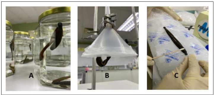
Figure 1: Leeches are placed in glass jar in starvation period (A), Leeches are sucking mixture of 0.001M Arginine and 0.1M NaCl solution (B), and Leeches are being gently massaged from posterior to anterior (C)
The relationship between total protein in CLSE and attachment period indicated that attachment period remarkably affects the total protein content. In other word, the longer the attachment period, the lower is the total protein concentration. The highest concentration was noticed after 2-5 mins of the sucking action started followed by a dramatic decline afterward as leeches were allowed to continue sucking the phagostimulatory solution.
The preliminary results for the first 14 days of the study found that the total protein concentration was very high compared to the following week throughout the study. In the following weeks, from the fourth week to the eighth week, there was a decrease in the concentration of total protein in the saliva collected from each group. Although the trend showed a decrease in the amount of protein, the amount of protein still existed until the eighth week of the study conducted (Table 1).
|
Group No. |
Starvation Period |
Total Protein Concentration (mg/ml ± SD) |
|
1 |
2 weeks |
49.8 ± 0.331 |
|
2 |
4 weeks |
40.34 ± 0.053 |
|
3 |
8 weeks |
24.45 ± 0.003 |
Whenever the starvation time was longer, the total protein activity, such as Hirudin, was lower. It was found that the starvation period is suggested to be no more than 16 weeks and can be extend to only 6 months [19]. LSE was less biologically active whenever leeches got exhausted and aged more than six months [19]. The time- dependent protein concentrations relationship could be referred to the more ingested volume of the phagostimulary solution, as leeches were allowed to stay attached for a longer time. The blood-sucking leeches were known to produce salivary secretions containing biologically active peptides and proteins, especially the antithrombin agent hirudin. One part of these secretions will be mixed with the sucked solution enabling the leech to store the meal for up to 18 months [20,21]. In the case of an artificial solution and upon feeding, leeches re-suck most of their saliva secretions into phagostimulatory [22]. This fact can explain the reason of the significant rise in protein concentration after a few minutes of attachment.
The results obtained from the disc diffusion test found no difference in the inhibition zone against S.aureus but showed the inhibition zone against E.coli. This shows that CLSE only has antimicrobial activity against E.coli. This is in agreement with previous studies by Abdualkader et al.
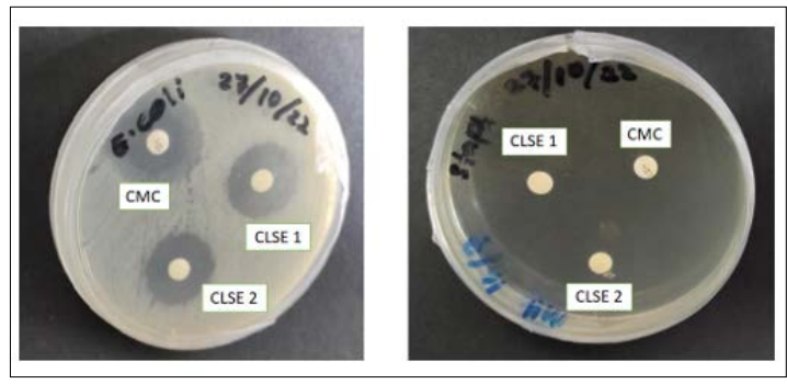
Figure 2: StaphylococousAureus and E.coli (CMC – Chloramphenicol as Positive Control, CLSE – Crude Leech Saliva Extract)
The result of MIC test is listed in Table 2. The CLSE possessed antibacterial activities against Salmonella enterica and Klebsiella pneumonia with a mean zone of inhibition ranged 20 – 45 mm. The test revealed that these bacteria were sensitive to the concentrations of 100% which ranged around 40 mm, and an inhibition zone in 25% concentration ranged 20 mm by Salmonella enterica. CLSE has revealed a spectrum of antibacterial activity similar to the result found by Abdualkader et al, S.aureus inoculum has no detected in microbial activity. Moreover, none of those bacteria exhibited any activity after a starvation longer than 28 days. The longer the starvation period, the less antimicrobial activity was found. As stated by Abdualkader et al, starvation period is a factor considered to determine the effect of antibacterial activity [13].
|
Medium |
Starvation Period (days) |
Bacterial species |
|||
|
Measurement zone (mm) |
|||||
|
E.coli |
Klebsiella pneumonia |
Salmonella enterica |
Staphylococcus aureus |
||
|
CLSE |
14 |
24 |
22 |
26 |
0 |
|
28 |
0 |
0 |
0 |
0 |
|
|
56 |
0 |
0 |
0 |
0 |
|
|
Ag + NaCl |
- |
0 |
0 |
0 |
0 |
|
CMC (50 mg/ml) |
- |
26 |
35 |
40 |
25 |
Reference Antibiotics 0: No Inhibition, CMC - Chloramphenicol
The finding suggests that the slightly breakdown of the nutrients results in a decrease in the amount of CLSE production which require less concentration of the protein to be secreted. A starvation period of more than six months is sufficient for most of the collected leeches to reach sexual maturity or even the end their life cycle [19]. Based on report from WHO, the widespred occurrence of antibiotic resistance suggests that, in principle, any organism could develop resistance to antibiotic drugs increase the cost of treatment and often results in treatment failure. The problem of microbial resistance is growing and the outlook for the use antimicrobial drugs in the future is still uncertain.
This study found that starvation period plays an important role in determining the production of CLSE. The production of protein concentration decreases during starvation even though there is still protein in the CLSE in a reduced volume quantity. The decrease in protein concentration after 4 weeks of starvation is believed to be related to exhaustion and aging. Antimicrobial activity was found in the growth of those organism tested. A further study is required to elucidate the CLSE before collection including leeches preparation in terms of of feeding, starving and extraction the CLSE. The CLSE collection method and treatment also needs to be taken consideration without affecting the quality and quantity of CLSE collection.
The authors would like to express their gratitude to Ministry of Science, Technology and Inovation (MOSTI), the Steinbeis Foundation and University College MAIWP International (PICOMS/RMC/Penyelidikan/2020 (04)) for their financial support.