Author(s): Nissrine AMRAOUI, Meriem MEZIANE, Salim GALLOUJ and Fatima Zahra Mernissi
Pregnancy is a physiologic condition with a significant alteration of the immune functions and during which extremities erysipelas are rarely reported. We report in the light of a literature review the results of 6 patients treated for leg erysipelas between 2012 and 2016.
Pregnancy is a physiologic condition with a significant alteration of the immune functions and higher risk of infection [1]. Erysipelas during pregnancy is rarely reported but severe forms occurred frequently [2,3,4]. About that, Cervicofacial location is the most common site reported [2]. The anatomy of the oral cavity plays a significant role in the spread of cervicofacial infection whose 75% is usually of odontogenic origin [5,6,7]. In this particular context, prompt management should be instituted to avoid fetal and maternal serious consequences and complications. Through this article, we attempted to raise in a series of six women the clinical and evolutionary characteristics of extremities erysipelas rarely reported in these locations during pregnancy.
This cohort descriptive study was carried out on 6 pregnant women with leg erysipelas to the dermatology department of Hassan II University Hospital (Fez, Morocco) within a four-year period from January 2013 to December 2016. Information was collected on the basis of patient case files, emergency registers as well as inpatient admission records. The study protocol was approved by our Institutional Ethics Committee. All patients benefited from obstetric examination to determine the pregnancy and fetal status.
The age of our patients ranged from 32 to 38 years with a mean age of 36. The term of pregnancy was respectively 32, 36, 33, 31, 28 and 8 weeks of amenorrhea (Table 1). All patients don’t have a secondary comorbid medical condition. The six patients had leg erysipelas without a previous episode. Of the six patients, four were obese. The entry point of the infection was a foot intertrigo in four cases, wound in one patient and excoriation secondary to pruritus in the last case. The clinical aspect was a well-defined, tender and bright red plaque, we also noted blisters in three cases, purpura and erosions in the one other patient (Figures 1 and 2). The Laboratory Risk Indicator for Necrotizing Fasciitis (LRINEC) score was 4 on average. The venous Doppler ultrasound did not show any abnormalities in all cases. In the present study, All the patients were reviewed by the obstetrics and gynecology team at the admission. The average time for consultation and hospitalization duration was 3 and 10 days respectively
| Patients | Age | Pregnancy term (Weeks of amenorrhea) | Number of pregnancies | Previous treatment | Management | Outcomes |
|---|---|---|---|---|---|---|
| 1 | 1 | 32 | 4 | traditional | Medical | Favorable |
| 2 | 38 | 36 | 3 | NSAIDs | Medico-surgical | Favorable |
| 3 | 36 | 31 | 4 | Unspecified | Medical | Favorable |
| 4 | 38 | 33 | 2 | NSAIDs | Medical | Favorable |
| 5 | 34 | 8 | 1 | Antibiotic | Medico-surgical | Favorable |
| 6 | 38 | 28 | 3 | Antibiotic | Medical | Favorable |
NSAIDs: Non-steroidal anti-inflammatory drugs
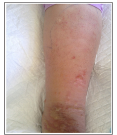
Figure 1: well-defined, tender, red plaque with blisters
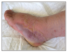
Figure 2: Red edema with Hemorrhagic bullae and erosions
Treatment used intravenous amoxicillin with clavulanic acid. The evolution was favorable in 4 cases, while two patients presented an abscess requiring the modification of antibiotic therapy to ceftriaxone and metronidazole, and they got surgical drainage. We also treated points of entry to avoid a recurrence. Fortunately, all pregnancies were completed without complication.
Pregnancy is a physiologic condition with a significant alteration of the immune functions [8]. In fact, Pregnant women are at a much higher risk of infection, compared to nonpregnant women because of a decrease of neutrophil chemotaxis, cell-mediated immunity and natural killer cell activity among them [5-11].
Extremities erysipelas is rarely reported during pregnancy, it is more usually described at cervico-facial location with a prevalence rate of 13.7% noted by Omeje KU et al. The authors consider that populations from poor and rural areas are more exposed to infection as well as its severe complications in the background of pregnancy [2].
In addition, some factors may play a major role in developing this infection including diabetes, obesity which was noted in four of our patients and important physiologic edema that is often observed at the last months of pregnancy due to hormone-induced sodium retention and inferior vena cava compression by an enlarged uterus [12,13]. In pregnancy, there is usually an upsurge in the levels of estrogen and progesterone. These hormones are largely responsible for marked skin changes and damages that promote infection hence the interest to use moisturizers [14,15].
Treatment includes first antibiotics that should be safe for use during pregnancy and effective against the bacteria causing erysipelas. About that, penicillin is the drug of choice [16,17,18]. tetracycline and aminoglycosides are often contraindicated in pregnancy due to their effect on teeth discoloration, developing bone and ototoxicity. As for metronidazole, it is relatively contraindicated due to its potential teratogenic effect [19]. However, it is administered when the benefit is deemed to outweigh the risk [19].
Uncontrolled comorbid medical conditions have been associated with poor prognosis in pregnant women. Osunde et al noted overwhelming sepsis in a pregnant patient with uncontrolled diabetes mellitus [17].
Poor initial management plays an important role in the occurrence of local erysipelas complications that occurred in 31% to 52% of patients [20,21,22]. In a total of 152 patients, the most frequent local complications were bullae (27%), followed by hemorrhagic lesions (20%), abscesses (11%) and necrosis (5%) [21]. In the case of Uncomplicated abscesses, the attitude consists of an incision and drainage. In this context, two recent trials showed that cure rate increases from 69-74% to 81-83% with adjunctive antibiotics [23,24].
From a bacteriological point of view, a study carried out by Thiebaut et al in 17 patients showed the frequency of aerobic germs in this type of infection. In our study, bacteriological examination isolated aerobic germs in 2 cases [25].
Lymphoedema and obesity are the most important risk factor for recurring erysipelas [26]. About that, all risk factors should be treated vigorously. When infections recur despite adequately treating risk factors, prophylactic antibiotics should be started [27].
Keefer and Spink reported a general mortality from erysipelas of 16.4 ± 3.6% prior to the use of antibiotics [28]. This mortality decreased with the advent of the chemotherapy to 2.4% according to Hoyne, Wolf and Prim [29].
According to Williams any septic condition has a more serious prognosis in pregnancy than at other times. Adair and Stieglitz, in their stated in their textbook “Obstetric Medicine,” that the prognosis of erysipelas rarely observed in pregnancy is not necessarily unfavorable, especially if the disease is limited. All of the patients in the present series had an improvement in their clinical status without systemic complications. The 2 patients who presented an abscess had benefited from surgical drainage with favorable evolution. Fortunately, all pregnancies were completed at term [30,31].
In pregnancy period, attention should be given to measures that prevent erysipelas by especially treating lymphoedema as well as all point of entry. Multidisciplinary and prompt management of erysipelas during pregnancy is important to ensure outcomes. Until recovery, close controls are necessary for the pregnant patient and the fetus to avoid complication particularly sepsis, preterm labor and premature birth.