Author(s): Anil Melath, Jilu Jessy Abraham*, Nanditha Chandran, Prithi S, Prudhvinath Chowdari E and Arjun MR
Suturing is necessary and important event in both oral and general surgeries [1]. The term ‘Suture’ describes any strand of material utilized to ligate blood vessels or approximate tissues without the necessity of a mechanical Support [2]. Moreover, Surgical Suture is nothing. but isolating the healing centre, promoting the circulation process, controlling haemostasis stabilising the tissues and the requested position. Various methods & materials used for precise flap placement [3, 4].
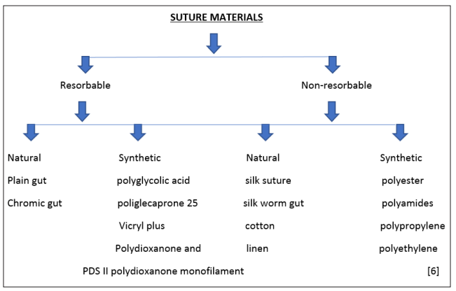
Suturing materials have important implications in tissue repair. A Suture should have high tensile Strength, knot security and easy to handle. Inadequate Suturing may result in flap skipping, exposed bone/necrosis, pain and delayed wound healing [5].
The aim of this study is to re-examine and to summarize the different Suture threads origin characteristics and their interaction on the tissues, to obtain better wound healing.
The first evident of Sutures goes way back to 30,000 BC where eyed needles were used for surgery and to tie wounds after the trauma according to fossilized remains of Neolithic skulls.
By the time 1600 BC Greek Surgeon, Galen of Pergamon noted that he has used Silk or Catgut (which is made from the twisted intestines of sheep or horses) to Suture together the gladiators who has injured skin, muscles and Tendons. Similar type of Sutures was used up to 20th Century.
The Egyptian records shows that the first historical evident of sutures is at 150 AD.
At 1887, the manufacturing of first used Sterile Sutures happened which were made up of either catgut or Silk.
At early 1920s, ‘Mer sutures’ were developed by Scottish Pharmacist ‘Merson’ which were eyeless needles with a single strand of material attached to the butt of the needle This method greatly reduces tissue damage by pulling two Strands through skin.
In 1969, remains ‘Polypropylene’ sutures were invented. Still as one of the best for cardiac bypass surgery. The usage is also easy.
In 1974, Vicryl Sutures were introduced into the market which are naturally absorbed into the skin. It is also strong and pliable.
In 1979, a coated version of Vicryl sutures were introduced. This has an advantage of knots sliding down more easily when a Suture is being tied.
In 1982, PDS II Sutures made Debut which were made of Polydioxanone which were designed to close fascia under the skin.
In 1993, Monocryl sutures were delivered to the market. This has a specific advantage of even more skin closure and prevents wound edges from separating. It has high initial strength.
In 1998, topical skin Adhesives were introduced. In 2003, coated Vicryl plus antibacterial Sutures were introduced [6].
Suture needle
Parts: Needle eye, Needle body, Needle point.
Needle eye
*Can be closed or swaged. Shape of eye may be round, oblong or square.
*They are traumatic and sometimes atraumatic.
*Expensive, but sterile [7]
Needle body
*Widest portion of needle
*Referred to as needle grasping area.
*Half circle curved needles are commonly used in oral surgical procedures [8].
Needle Point.
*Extreme tip of the needle, it can be cutting, round or blunt.
*Blunt point has rounded end and is useful in friable tissue suturing like parotid/lacrimal duct, etc.
*Round/tapered needles used for suturing soft and non-keratinized tissues such as muscle, fascia and neural sheath [9].

The main aim of a Suture is to minimise or eliminate dead space in skin and sub cutaneous tissues. Lower the tension that causes wound separation. It also involves with correct wound placement in order to relax tension lines. The technique which is used to close the traumatized area depends on the force and direction of tensions of wound, The thickness of the tissue and anatomic considerations. The types mentioned below
Knot tying is significant to tie knots while using monofilament nylon sutures because of the risk of Separating out. If the knot starts to slip after the first two throws by increasing the frictional force needed to unravel the knot by pulling it in the corresponding direction rather than pulling it in 90° [10-12].
Simple interrupted Suture is the most popular suturing technique of wound closure used in cutaneous surgery. To place a simple interrupted suture, The needle enters one side of the wound and penetrates well into the dermis of the sub cutaneous tissue. This technique can be used for the wounds with uneven thickness by changing the angle and the depth of needle used. Then the needle is passed through the subcutaneous tissue to the opposite site of the wound and is placed closer to the wound edge • So that the final configuration of the suture is flask shaped.
Vertical Mattress suture is the best available suturing techniques to ensure of wound and to reduce the wound tension. It is started at 0.5-10cm lateral to the wound Margin. It is inserted to the depth of the wound ensuring the closure of dead space. The needle is then passed to the deep tissue to the opposing wound edge, where it comes out of the skin on the opposing side equidistant to the insertion. It is then reversed and the skin is penetrated again on the side where the suture just excited but closer to the wound edge.
Near Far vertical Mattress Suture is a modification of standard vertical mattress suture [10,13]. It should begin near the wound edge (1-3mm) Passing needle into the deeper aspect of the opposing side and exit through the epidermis wide to the insertion (0.5- 1.0cm). Reverse the needle and re-entry skin near the wound edge (1-3mm) of the side just excited and repeat the same procedure exciting wide to the initial preparation. It is desirable for tissue expansion and wound closure which is under tension.
Horizontal Mattress suturing technique helps in minimizing the wound tension, closing the dead space and facilitating wound edge eversion [10-13]. It Penetrates the skin 5-10mm from the edge of the wound the needle is then passed dermally or Subcutaneously towards the opposing wound edge where it enters at the same level in the Subcutaneous or dermal tissue. Exit the opposing side of the wound through the epidermis at equidistant. Re-entry the skin on the same side at the sooner distance from the wound edge but laterally. The needle is then passed dermally to the side of former preparation.
Running suture, this technique is useful over eyelids ears and the dorsa of hands or can be used to secure the edges of full or split thickness skin graft [10-13]. The primary advantage of this technique is its relative care and speed of placement. Initially place a Simple intercepted Suture at one end of the wound. This is tied but is not cut. Simple sutures are placed down the length of the wound, repenetrating the epidermis and passing dermally or subcutaneously. It is important to space each interval of the running Suture entirely. It is terminated by placing a single knot as it exists the skin at end of incision.
There are also different types which includes:
A granny knot consists of two simple throws, with one throw beginning with the instrument between each strand of suture and the other beginning with the instrument outside of the strand of suture. Hold two ends of the laces and that should be joined together and hold them. Put the leftover the right lace, similar to an overhand knot. Pull the ends of the laces towards one another. Then pull the left lace over the right place. Finally, pull both ends and make sure that it is strengthened.
The main advantages of the Granny knot are that it is quite easy to tie. Then, it is the basis of the surgeon’s knot, which makes it widely used by surgeons.
The disadvantages of the Granny knot are related mainly to its strength It is not so secure as other knots, can easily become undone and may even slip when they are overloaded. When there is an excessive tightening of the Granny knot it might jam, so it is better to consider it prior to using it for holding heavy weights.
Ethicon (1985) recommends the following principles for knot tying:
Sutures should be removed as a traumatically and cleanly as possible. Ethicon (1985) recommends the following principles for Suture Removal:
In 1998 topical skin adhesives were introduced.
In 2003 coated Vicryl plus antibacterial sutures are introduced. coated Vicryl plus antibacterial sutures has antibacterial property as the name indicates.
In 2006, INSORB, subcutaneous absorbable stapling device, became commercially available In 2009 Covidien introduced V-LOC unidirectional barbed suture with a first loop. Ethicon has now introduced its own version of the barbed suture, the Stratafix found in both unidirectional and bidirectional.
In 2011,2012 and 2014 Ever point cardiovascular needles, Stratafix knot less tissue coated devices and Dermabond prineo skin closure systems were introduced respectively [17-19].
Management of soft tissues is a supreme Priority for surgeon in any of the extra or intra oral surgical or invasive surgical procedures for aesthetics. Sutures not only perform their primary function of wound closure but they afford other benefits such as antimicrobial properties and more recently elimination of the surgical knot with barbed suture and replacement of skin sutures with adhesions Proper soft tissue handling during various suturing techniques can ensure optimal tissue healing and high aesthetic result [20,21]. The success of technique sensitive surgeries depends on the clinician’s knowledge and skills to close the wound and achieve optimal healing. The innovation in suturing materials decreases the potential for postoperative infections [22-26].
Conflict of interest: Nil
