Author(s): Amani Sastry and Victor M Jimenez*
Toxoplasma gondii (T. gondii) is one of the most common parasites that infect humans. The parasite exists in approximately 40 million people in the United States. Latent infections are frequently associated with tissue cysts of T. gondii in the skeletal muscle and brain tissue that can lead to mental disorders, congenital disorders, and vision dysfunction. Furthermore, self-directed violence, impulsivity, and aggression are associated with T. gondii infection. Dopamine is associated with human behaviors including pleasure, aggression, memory, and substance use disorder; however, the studies on the association of T. gondii infection and drugs of abuse are not well understood. The frequency of substance use may be associated with substance-induced modification of dopamine-receptor densities and basal dopaminergic activity. Likewise, T. gondii can directly or indirectly influence dopaminergic activity in infected cells. The current scoping review aimed to review peer-reviewed literature and identify gaps to evaluate the association between drugs of abuse and T. gondii infection. Articles for review were included if published between the 1st of January 1970 and to the 30th of December 2022. Different key words and phrases were adopted: “T. gondii infection”, “Toxoplasmosis”, “T. gondii and drug use”, “T. gondii infection and dopamine”, “T. gondii infection and drugs of abuse”, “drugs of abuse”, “T. gondii infection and alcohol”, “SUD”, “Toxoplasmosis and SUD” and “Substance abuse and inflammation”. Articles that did not match and/or were published outside of history range were excluded. Databases utilized to generate results included PubMed, Web of Science, and Scopus. One hundred and nine articles or resources were generated and deemed appropriate for reference in this review. Results indicate that individuals infected with T. gondii display increased risky behavior, such as excessive alcohol consumption. T. gondii seropositive subjects had a reduced likelihood of self-reported substance use compared to T. gondii seronegative subjects which confirms that SUD is a potential risk factor for behavioral and psychiatric complications associated with T. gondii infection. Therefore, it is necessary to conduct further research characterizing the mechanisms associated with dopamine metabolism of drug dependence and withdrawal in the context of a T. gondii infection, evaluating the role of inflammation, and identifying potential drug-and-sex specific underpinnings of these associations.
Toxoplasma gondii (T. gondii) is an obligate intracellular parasite that infects approximately one-third of the human population. Members of the feline species are definitive hosts of T. gondii while humans are intermediate hosts [1]. Toxoplasmosis is more commonly presented as a latent infection whereas those infected are asymptomatic; however, severe symptomatic disease may precipitate in immunocompromised individuals and congenitally infected fetuses [2]. Symptomatic T. gondii infection may present as lymphadenopathy, chorioretinitis, hydrocephalus, and mental status changes [2]. Toxoplasmosis of immunocompromised individuals, such as acquired immunodeficiency syndrome (AIDS) patients, is often a result of reactivation of latent infection ultimately presenting as neurological dysfunctions [3]. For example, an increased frequency of Toxoplasma encephalitis in AIDS patients is recorded, specifically those with CD4+ helper T cell counts less than 200 cells/µL [4].
Of the myriad clinical presentations in individuals infected withT.gondii, the neuropsychiatric findings and behavioral changes associated with this parasite continue to be an area of research not well understood. Recent studies have suggested that latent T. gondii infection is not entirely asymptomatic, with evidence of behavioral alterations in both rodents and humans infected with T. gondii such as increased impulsivity, aggression, risky behavior, and suicidal behavior [5-7]. Furthermore, many studies have found higher incidence of T. gondii infection in schizophrenia patients as well as anxiety, depression, and other mental health illnesses [8,9]. Although evidence for T. gondii infection in the context of drug dependence and the development of substance use disorder (SUD) are variable, the biological link to T. gondii’s dormant stages in the brain, subsequent neuroinflammation and dopamine metabolism, remain to be elucidated. The current scoping review will examine the following objectives: 1) identify T. gondii’s mechanism of pathogenesis, 2) identify the mechanism of action for prevalent drugs of abuse, and 3) identify links between drugs of abuse and T. gondii infection in the literature. In addition, any gaps in the literature will be identified and reported. The current review examines the neurological impact of drugs of abuse and T. gondii infection. Literature evaluation of the overlapping impact on the brain as a centralized source for injury, associated with neurobiological, cognitive, neuropsychiatric, and behavior pathologies can improve targeted treatment strategies for toxoplasmosis. Beyond treatment strategies, the current research findings will improve clinicians and researchers understanding of the etiology and immuno-pathophysiology of related diseases and facilitate improved patient outcomes.
The scoping review was guided by the Systematic Reviews and Meta-Analyses -Extension for Scoping Reviews (PRISMA-ScR). The completed PRISMA ScR Checklist is provided as File 1 in the Supporting Information. Scoping reviews are a type of review which follow a systematic approach to map evidence on a topic and identify main concepts, theories, sources, and knowledge gaps. A review protocol is not registered.
Scientific journal research articles, study designs, reviews, reports, book chapters, conference presentations, and publications were included if deemed relevant to the central research question. Publications published between 1970 and end of year 2022 had to refer to T. gondii infection and any associated determinants such as drugs, substance use disorder, or conditions of inflammation. In addition, studies exploring specific drugs of abuse, neurological or behavioral implications were also included. The setting included the United States and international data.
The included articles were searched on the electronic databases, PubMed, Google Scholar, Web of Science, and Scopus. These were identified as the most relevant databases where all types of studies on T. gondii and drugs of abuse could be located. Different key words and phrases were adopted: “T. gondii infection”, “Toxoplasmosis”, “T. gondii and drug use”, “T. gondii infection and dopamine”, “T. gondii infection and drugs of abuse”, “drugs of abuse”, “T. gondii infection and alcohol”, “SUD”, “Toxoplasmosis and SUD” and “Substance abuse and inflammation”. Articles that did not match and/or were published outside of history range were excluded. The keywords were matched through the Boolean operators AND OR in the databases.
Two researchers independently reviewed all the titles, abstracts and full texts in a stepwise process using the eligibility criteria described previously. The article or source was included if there was agreement between the researchers. If conflicting agreement, a third researcher arbitrated. Publications written in the English language were reviewed.
Synthesis of the content was guided by the study aims and each aim was analysed in terms of key findings, successes and enablers, challenges, and barriers to T. gondii and drugs of abuse, gaps in the evidence and further research required. A thematic analysis was conducted and is presented along the following main deductive themes: T. gondii infection, neurological inflammation, substance abuse and inflammation, drugs of abuse and T. gondii, further evidence linking T. gondii infection and alcohol use disorder, further evidence linking T. gondii infection and psychiatric drugs. In addition, the first author and year of publication, the objective of the study, the conclusion of the study, population screened, and main results were stored. A description and narrative synthesis were adopted to describe the results.
T.gondii is an obligate intracellular parasite transmitted primarily through the consumption of oocyst contaminated foods [10]. Feline species are definitive hosts of T. gondii and can shed highly infectious oocysts into the environment generating a wide variety of intermediate hosts [10]. The three main stages in its life cycle are the following: sporozoites (oocysts), tachyzoites (replicative stage), and bradyzoites (tissue cysts) [10]. Once the environmentally stable oocysts have been ingested by the intermediate host, the parasite transitions to the acute stage of infection [10]. This stage involves rapidly replicating tachyzoites that disseminate throughout the body. Tachyzoites replicate every 6 to 8 hours within an intracellular parasitophorous vacuole (PV) which is essential to the growth of the parasite [11]. T. gondii infection transitions to the bradyzoite stage, a slower replicating form, characterized by the presence of tissue cysts that lie dormant in muscle and brain tissue marking chronic infection with the parasite [10]. Immuno-competent individuals with acquired toxoplasmosis are generally asymptomatic and are protected from symptomatic re-infection by host immune defenses. In contrast, immunocompromised individuals with chronic infection, such as patients with AIDS, can experience reactivation of the latent infection in the brain, otherwise known as cerebral toxoplasmosis, and other organs where tissue cysts lie dormant [10]. Reactivation of latent infection in the brain can be life threatening as bradyzoites convert to actively replicating tachyzoites resulting in Toxoplasma encephalitis (TE). Acute TE is a result of the parasite infecting microglia, astrocytes, and neurons and then later persisting in mainly neurons, marking chronic TE [12]. During the transition from acute to chronic TE, the CD4+ and CD8+ T cell levels gradually decline, with CD8+ T cells at very low levels during chronic TE [13]. Therefore, to understand the complexity of T. gondii infection in both immuno-competent and immuno-compromised individuals, the characteristics of acute versus chronic infection and the specific mechanisms T. gondii utilizes to manipulate host immune responses must be identified (Figure 1).
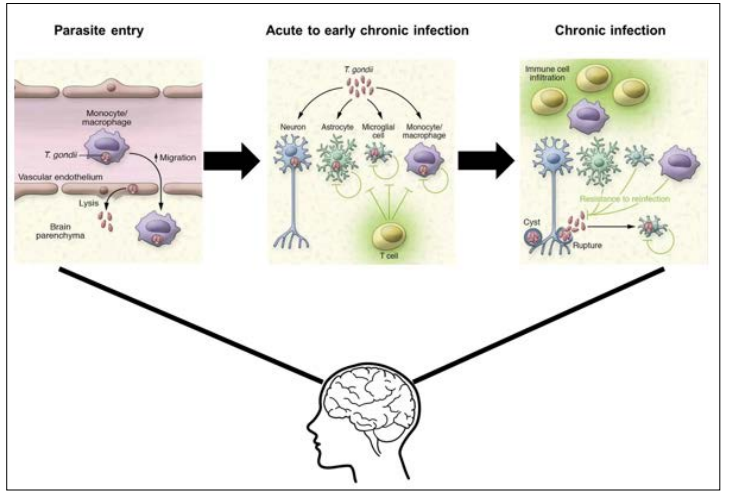
Figure 1: Acute T. gondii infection begins with infection of immune cells and then subsequent lysing of the brain vascular endothelium due to increased blood brain barrier (BBB) permeability. In the transition to early chronic infection, the parasite infects neurons, astrocytes, microglia, and monocytes eliciting an immune response. Chronic infection is characterized by the presence of tissue cysts in neurons and other immune cells that lie dormant and are controlled by host immune response. Image modified from Zhao and Ewald, 2020.
Diagnosis of toxoplasmosis relies on the presence of specific immunoglobulin M and G (IgM and IgG) antibodies to Toxoplasma antigens. Acute toxoplasmosis can be identified by the presence of Toxoplasma-specific IgM antibodies; however, it has been reported that IgM may last longer in the serum following infection [14]. Therefore, the IgG avidity test determines the timeline of acquired infection based on IgG affinity, with low avidity IgG antibodies indicating acute infection [14]. Transmission can also occur congenitally during acute toxoplasmosis in a seronegative mother. Tachyzoites present in the blood may cross the placenta and infect the fetus leading to serious complications and birth defects [10]. Conclusively, T. gondii infection can be narrowed down to acute and chronic infection. Acute infection is characterized by actively replicating tachyzoites that, in immuno- competent individuals, can be cleared by host immune defenses. The parasites that do survive persist as slow-growing bradyzoite tissue cysts in tissues such as the brain, eye, cardiac, and skeletal muscle [15]. Chronic infection is still poorly understood but can be characterized by the sustained immune response to T. gondii as illustrated by the elevated Toxoplasma-specific IgG and IFN-γ in the sera [16]. Less well understood is the immunological response to infection. Nevertheless, T. gondii infection relies on the manipulation of host immunity through the secretion of specific immunoregulatory cytokines, control of gene transcription, and modulation of cell adhesion and migration pathways. The type 1 T helper (Th4) type cell-mediated immune response is the primary resistance mechanism to the T. gondii infection. This response involves the production of IL-12 by dendritic cells, macrophages, and neutrophils that then stimulate natural killer cells and T lymphocytes to produce IFN-γ [17,18]. IFN-γ plays a major role in controlling both acute and chronic stages of infection, by activating anti-parasitic effector cells such as macrophages, fibroblasts, and astrocytes [19]. For immunocompetent individuals, the host adaptive immune response is imperative for the control of replication during an acute infection and the eventual formation of cysts in the brain and other organs that persist throughout the host’s life. Within a few weeks following acute infection, the host immune response eliminates tachyzoites. However, in immunocompromised individuals this process is hindered by the reduced CD4+ T cells population, leading to a decrease in IFN-γ levels and subsequent chronic infection in the brain [20]. Chronic T.gondii infection is characterized by the prevalence of bradyzoites
and tissue cysts. These bradyzoites can be found in the brain as early as 3 days following infection and mature into tissue cysts primarily located in the grey matter and neurons of the brain [10]. CD8+ T cells are key adaptive immune system mediators for the control of chronic infection and preventing reactivated infection in the brain. CD8+ T cells stimulate production of pro-inflammatory cytokines, predominantly IFN-γ, as well as anti-inflammatory cytokines, such as IL-17 and IL-27 [20]. Activated CD8+ T cells have also been shown to lyse Toxoplasma-infected cells through perforin-mediated cytotoxic activity [21]. A recent study performed by Suzuki et. al. suggests that this perforin-mediated cytotoxic activity by CD8+ T cells was able to remove Toxoplasma cysts from the brain [21]. IL-10 inhibits the production of IL-12, IFN-γ, TNF- α, and IL-6 from macrophages and microglia in the brain, thus limiting the lethal inflammatory response that causes TE [22]. Ultimately, understanding the key players involved in host immune response is crucial for controlling T. gondii infection and can provide new insights for potential therapeutic targets to reduce disease.
Additionally, tachyzoite to bradyzoite development in T. gondii is a crucial aspect of the parasite’s life cycle, specifically with the formation of tissue cysts, and can be attributed to epigenetic and transcriptional regulation [23]. The factors contributing to the tachyzoite transition into the bradyzoite stage are still poorly understood. However, the transition is associated with cell stressors, such as oxidative stress or heat shock, and the transcription of bradyzoite-specific proteins necessary for tissue cyst formation [23,24]. For instance, a group of 67 ApiAP2 transcription factors (a family of DNA binding proteins) are in the Toxoplasma genome; 24 proteins are expressed during the tachyzoite replicative cycle and several of the same factors are also implicated during bradyzoite development [23]. The specific ApiAP2 factor, AP2IV-4, was shown to be expressed during the tachyzoite cell cycle; however, upon deletion of the locus, there was subsequent expression of bradyzoite-specific proteins in the replicating tachyzoites [23]. Ultimately, these data suggest that the mistiming of bradyzoite antigens in tachyzoites lacking AP2IV-4 caused a potent inflammatory monocyte immune response that effectively eliminated this parasite and prevented tissue cyst formation in mouse brain tissue. Bohne et al observed that IFN-γ treatment induced bradyzoite gene expression in tachyzoite- infected mouse macrophages but not in fibroblasts, suggesting cell type-specificity [25]. If the host is immuno-compromised, T. gondii shifts toward its tachyzoite replicative form in a process called recrudescence which is associated with tissue damage from a CD4+ T-cell mediated immune response (Figure 2) [26].
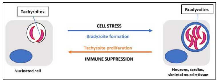
Figure 2: T. gondii in its tachyzoite form will invade and replicate within any nucleated cell forming a parasitophorous vacuole. During infection, cell stressors such as changes in pH or oxidative stress can promote bradyzoite differentiation in predominantly neuronal, cardiac, or skeletal muscle tissue.
Virulence is influenced by the genotype of the parasite. Type I strains are considered highly virulent in mice compared to Type II and III strains, which are more commonly associated with human toxoplasmosis [27]. Type I strains are characterized by high motility in vitro allowing for easier transmigration and dissemination whereas type II strains rely on host cell trafficking. During infection, the control of toxoplasmosis is characterized by an elevated IFN-γ dependent Th4 cytokine response. Type I and Type II T. gondii strains, elevate IFN-γ, TNF- α, IL-12, and IL-18 expression during lethal infection [11]. Type II strains have been shown to induce stronger proinflammatory responses, with a prominent expression of IL-12, compared to types I or III. Structurally, T. gondii at its invasive tachyzoite stage contain highly polarized cells and specialized secretory organelles called micronemes, rhoptries, and dense granules, which secrete virulence factors responsible for dissemination and survival of the parasite. Exocytosis of these secretory organelles mediates the formation of a PV which protects the parasite from the host cell and provides nutrient sources for development. Additionally, the rhoptries and dense granules secrete effector proteins as defense mechanisms against host cell responses. These effectors are highly polymorphic and differ based on T. gondii strain [27]. These polymorphic differences illustrate the complex nature of strain- specific virulence and host-pathogen interactions that allow T. gondii to disseminate within its host. Ultimately, the expression of specific genes and virulence are important characteristics of this parasite’s life cycle and can be tracked via the associated immune response.
Parasite egression is another key component of T. gondii infection and dissemination into the brain. One mechanism is through infection of leukocytes as a vehicle for transport into target organs. For instance, T. gondii has been shown to infect host macrophages thus causing a suppression of proinflammatory cytokines, such as IL-12 and TNF- α. This is accomplished by manipulation of the signaling cascade, Nuclear Factor- kappa B (NF-kB), specifically through the activation of STAT3 [28]. The parasite can then cross capillary walls of the target organ by infecting endothelial cells. In a study conducted by Baba et. al., it was revealed that adhesion of T. gondii tachyzoite infected leukocytes to endothelial cells triggered immediate egression of the parasite into the target organ, thus preventing the opportunity for an immune attack [29]. The mechanism of parasite egress is a crucial aspect of T. gondii infection and warrants further research into the specific cytokine expression during the transition from acute to chronic stage. Furthermore, behavioral manipulation is a key component of T. gondii infection which may be attributed to alterations in dopamine metabolism; however, the mechanism for these changes is currently unknown. In a previous study conducted by Stibbs et al, it was observed that dopamine levels were 14% higher in mice with chronic T. gondii infection compared to controls, while other neurotransmitters were unchanged [30]. Subsequently, recent studies have identified two possible mechanisms that may play a role in behavior manipulation, 1) alteration of neurotransmitter signal transduction, indicated by the cessation of parasite-induced behavioral changes when administered medications for psychiatric diseases, and 2) the presence of tyrosine hydroxylase encoded in the parasite’s genome [31]. Regarding psychiatric disease,T.gondii seroprevalence has been increasingly associated with schizophrenia. Dopamine dysregulation has been shown to play a significant role in the development of schizophrenia which is why the principal antipsychotic medication used in its treatment is dopamine antagonist, haloperidol [32]. Webster et al found that haloperidol as well as valproic acid, a mood stabilizing medication also used in the treatment of schizophrenia, prevented the development of behavior changes in latent toxoplasmosis- infected rodents [33]. Furthermore, tyrosine hydroxylase is a rate-limiting enzyme involved in dopamine synthesis and induced during differentiation of the parasite’s tissue cyst stages. A study conducted by Prandovszky et al found tyrosine hydroxylase present in intracellular brain tissue cysts as well as a three-fold increase in dopamine release from T. gondii infected dopaminergic cells compared to uninfected cells in vitro [34]. Ultimately the presence of this enzyme may be responsible for the increased dopamine levels observed during infection. With the understanding that in brain tissue, T. gondii cysts contain hundreds of bradyzoites, it is plausible that chronic T. gondii infection could enhance dopamine release in vivo. Taken together, dysregulation of dopamine metabolism can cause serious consequences on human behavior and potentially lead to neurological or psychiatric disorders, making it crucial to examine whether T. gondii infection directly or indirectly alters dopamine levels in the brain (Figure 3).
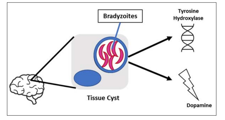
Figure 3: Tissue cysts localized in the brain enhance dopamine production, however the mechanism is still unclear. The presence of tyrosine hydroxylase, a rate limiting enzyme involved in dopamine synthesis, encoded in the parasite’s genome may be a contributor to the mechanism
The innate immune response of the central nervous system consists of resident macrophages, known as microglia, as well as mast cells, astrocytes, and neurons. These microglia serve as primary immune cells in the brain and spinal cord with the functions of phagocytosis and activation of cytokines [35]. Chronic T. gondii infection is characterized by the presence of dormant bradyzoite cysts localized in muscle tissue and more dangerously, the brain. Toxoplasma encephalitis (TE) is caused by the reactivation of these bradyzoite cysts into fast-replicating tachyzoites resulting in serious brain damage, especially for immunocompromised individuals [36]. However, it is still unclear how T. gondii enters the brain and evades the host immune system. Recent reports suggest that tachyzoites disrupt the host leukocytes’ natural migration phenotype, such as dendritic cells, and disseminate systemically through a “Trojan Horse” mechanism to rapidly reach the brain [37,38]. The “Trojan Horse” mechanism is characterized by T. gondii tachyzoites that infect host leukocytes and cross the blood brain barrier without being detected [39-42]. Moreover, Konradt et al observed that tachyzoites invade and lyse endothelial cells in brain vasculature prior to dissemination in the central nervous system [43]. In addition to leukocytes, studies test the role of glial cells for parasite migration and dissemination. Dellacasa- Lindberg et al showed that Toxoplasma-infected microglia exhibited minimal upregulation of major histocompatibility complex (MHC) II molecules and the costimulatory molecule, CD86, as well as a dramatic migratory phenotype when infected with live intracellular tachyzoites [44]. However, when studying IFN-γ stimulated microglia, this migratory phenotype was minimal in comparison to the tachyzoite-infected microglia [44]. Therefore, which cytokines are expressed that promote parasite migration and entry into the brain constitutes a noteworthy area of research. Hwang et al. showed an elevated and maintained expression of IL-12 and TGF-B for 12 weeks post infection as well as activation of the M1-type microglia, indicating a mixed anti-inflammatory and pro-inflammatory immune response for the control of infection [45]. Taken together, these studies suggest that parasite migration and dissemination require tachyzoite infection of host leukocytes and glial cells; however, further research must be conducted to understand the specific cytokine profile involved which may elucidate how T. gondii evades the host immune response upon entry to the brain, thus providing foundational knowledge for future therapies to combat the disease.
Substance use has highly stimulating effects in the central nervous system that could lead to drug addiction and serious health complications, such as memory loss and cognitive decline. These conditions are primarily associated with neuroinflammation and neurotoxicity that cause neuronal cell damage. Recent research has shown that drugs of abuse disrupt the BBB by altering tight junction formation making the brain susceptible to neuroinflammation [46]. Alternatively, the neuroinflammation resulting from exposure to drugs of abuse can disrupt the BBB further increasing its permeability to foreign toxins. An important aspect of the innate immune response and regulation of inflammation is the transcription factor, nuclear factor kappa light chain enhancer of activated B cells (NFkB) [47]. NFkB activation, upon exposure to drugs of abuse, causes innate immune gene induction leading to further inflammation and a progressive increase in addiction [48]. In the following sections, drugs of abuse and their effects on inflammation will be reviewed.
The neuropharmacology of amphetamines enhances the release of dopamine and serotonin as well as glutamate into the CNS. However, chronic amphetamine use can result in neurotoxicity marked by activated microglia [49]. Microglial activation is also involved in methamphetamine-induced neurotoxicity, specifically in the striatum, cortex, and hippocampus [50-52]. Subsequently, the activation of microglia results in secretion of proinflammatory cytokines and other reactive species that can cause damage to neuronal tissue. There is limited research on the specific inflammatory mediators involved, but Orio et al was able to show a significant increase in IL-1b after administration of MDMA [49]. Therefore, neuroinflammation following amphetamine-related drugs of abuse can be noted by the increase in activation of microglia and subsequent secretion of proinflammatory cytokines as well as oxidative stress. Likewise, cocaine use activates astrocytes through signaling of innate immune receptors and subsequent upregulation of proinflammatory cytokines, such as IL-1b. For example, Brown et al observed that chronic cocaine use activated innate immune signaling pathways that ultimately contribute to the mechanisms of cocaine seeking [53]. Furthermore, chronic cocaine abuse is implicated in HIV-1 associated neurological complications. This is illustrated by the enhancement of HIV-1 replication, increased permeability of the BBB, and the secretion of cytokines that decline CD4+ T cell counts. For example, Barr et al demonstrated that acute cocaine administration increased BBB permeability to the tracer, sodium fluorescein [54]. Regarding cytokine expression, it was recently shown that active cocaine users expressed higher levels of IL-6 and downregulation of IL-10, an anti-inflammatory cytokine [54]. Conversely, it was shown that active cocaine users have decreased IL-6 and TNF- α expression [54]. Therefore, further research must be conducted to understand the inflammatory response upon psychostimulant exposure as it may provide context for the development of severe disease in immunocompromised individuals.
Alcohol use induces variable effects on neuroinflammation responses that ultimately causes neurodegeneration. For example, Crews et al observed that ethanol-induced NF-kB activation mediates the neurotoxicity caused by in vivo models of binge ethanol administration [55]. Specifically, alcohol treatment of brain slice cultures resulted in increased NF-kB binding to DNA markers in gene promoter regions and decreased protective transcription factor binding [55]. These specific transcription factors promote neuronal survival and protection from excitotoxicity. The increase in NF-kB transcription results in upregulation of proinflammatory genes in the brain and less protection leaving the brain susceptible to damage [55]. Chronic alcohol exposure increases proinflammatory gene expression through activation of toll-like receptor 4 (TLR4), however acute exposure has shown opposite results. For example, Pruett et al showed a weakened TNF-α, IL-1β, and IL-6 immune response to LPS upon acute ethanol exposure in mice [56]. As for chronic administration, one study showed a marked increase in proinflammatory gene induction upon binge treatment with alcohol for 10 days in mice followed by LPS once the alcohol was cleared from system [57]. In addition to cytokine expression, microglia activation is another marker of alcohol’s role in facilitating inflammation. It has been shown that alcohol can activate microglia directly through the stimulation of TLRs or indirectly from release of damage- associated molecular patterns [58,59]. Ultimately, the stimulation of these microglia is characterized by proinflammatory cytokine expression, oxidative stress, and further neuronal damage. Taken together, alcohol both acute and chronic administration influence neuroinflammation which increases neurodegeneration and the potential development of chronic illness.
Opioid use is primarily associated with pain management; however, it is also implicated in the manipulation of inflammatory pathways. Like cocaine, opioid abusers have worse overall health and have an increased susceptibility to infectious disease, such as HIV infection. This is illustrated by immunosuppression upon opioid exposure such as decreased activation and chemotaxis of leukocytes and granulocytes upon morphine administration [60]. Regarding cytokine expression, there is variation as to which cytokines are upregulated and downregulated. Piepenbrink et al has shown heroin users have higher levels of inflammatory cytokines such as TNF-a and IL-8 [61]. However, Meijerink et al observed heroin abusers have lower levels of proinflammatory cytokines after the immune cells were in vitro stimulated by bacterial endotoxin lipopolysaccharide (LPS) [62]. Furthermore, acute, and chronic administration of morphine produces mixed cytokine expression as shown in various rodent models. In both acute and chronic administration there was an increase in peripheral IL-1b expression as well as in CNS preparations, including the NAc [63]. Additionally, there was an increase in IL-6 and IL-17 upon chronic administration and an increase in IL-10, TNF-a, IL-2, and IFN-c upon acute administration [64-66]. This finding is indicative of the immunosuppressive properties of opioid use as well as an increased risk of developing chronic illness.
The inflammatory effects of hallucinogens or psychedelics are still not well understood, but recent studies have suggested anti-inflammatory and immune-modulating properties of these substances. For example, LSD has shown the ability to suppress the proliferation of B cells and the production of pro-inflammatory cytokines IL-2, IL-4, and IL-6 in in vitro splenic lymphocytes derived from female rats [67]. Additionally, a human study looking at ayahuasca effects in healthy volunteers observed a decrease in CD4 and CD3 cells and an increase in natural killer (NK) cells compared to placebo group and subjects treated with D-amphetamine [68]. Furthermore, one study observed suppression of pro-inflammatory cytokine production upon administration of harmine, a component of ayahuasca, in mice treated with LPS simulating acute kidney injury [69]. Ultimately, these studies suggest that these substances may have therapeutic effects for autoimmune diseases, but there is still a need for deeper investigation of these drugs’ effects on the immune system.
Marijuana, otherwise known as cannabis, contains cannabinoid compounds that act on various receptors involved in regulating the immune response. This can be exemplified by anti-inflammatory effects because of decreased release of pro-inflammatory mediators [70]. The specific receptor proteins, CB1 and CB2, interact with cannabidiol (CBD) to provide anti-inflammatory and antioxidant properties [70]. However, it is important to distinguish that CBD is the modulator of these properties as opposed to THC which does not affect the serum levels of pro-inflammatory cytokines [71]. The role of the endocannabinoid system regarding neuroinflammation is exemplified by the increased expression of CB1 receptor and the potential upregulation of CB2 receptors upon neuroinflammatory conditions in the brain [70]. A study observing the cytokine expression in rats’ brain upon treatment with LPS showed a decrease of TNF-α, IL-6, IL-1b, and IL-12 levels when cannabinoids were administered [70]. The study also showed an increase in the production of TNF-α, IL-6, IL-1b, and IL-10 when cannabinoids were administered alone [72]. This data shows that the endocannabinoid system has conflicting effects with cytokine expression, but there is a significant impact on their production and function. Additionally, the presence of TNF-α induces expression of CB1 and CB2 which is partially a result of activating NF-kB which triggers transcription of inflammatory proteins and cannabinoid receptors [73] (Figure 4).
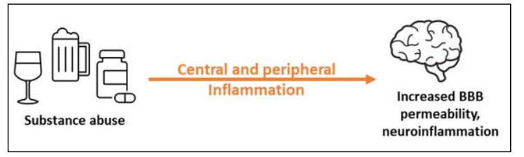
Figure 4: Substance use, such as alcohol, have inflammatory effects that may be implicated in increased BBB permeability and subsequent neurotoxicity when abused
Drug abuse, a devastating and harmful neuropsychiatric disorder, continues to be a widely concerned public health issue. Drugs of abuse, such as alcohol, cocaine/psychostimulants, hallucinogens, opioids, and marijuana, all contribute to neurotoxicity and inflammation leading to further complications in the brain. In this section, we will review each drug of abuse to describe their mechanism of action and examine biological connections with T.gondii infection.
Cocaine and psychostimulants, including amphetamines, methamphetamines, MDMA, etc. are sympathomimetics that inhibit catecholamine reuptake thus causing increases in blood pressure, heart rate, and body temperature. Abuse of these drugs can result in severity of these symptoms and possible death. According to the National Center for Health Statistics (NCHS) at the Centers for Disease Control and Prevention (CDC), the number of deaths since 2014 involving psychostimulants has significantly increased each year with 23,837 deaths in 2020 [74]. Cocaine related death has also increased since 2014 with 19,447 deaths report in 2020 [74]. These drugs act on the brain by increasing the availability of norepinephrine, dopamine, and serotonin at the synapse thus changing behavior and providing a sense of euphoria, alertness, agitation, and hyperactivity. However, more dangerous side effects of cocaine and psychostimulant abuse involve paranoia, psychosis, and hallucinations. Additionally, the inhibition of dopamine and monoamine reuptake can result in imbalanced free radical accumulation leading to oxidative stress and neuroinflammation. This neuroinflammation can be illustrated by activation of microglia and other inflammatory mediators that increase blood brain barrier permeability thus leading to loss of integrity and susceptibility to peripheral toxins in the brain. Likewise, T. gondii infection has been shown to alter neurotransmitter transmission either directly or indirectly. The direct effect can be illustrated by the presence of cysts in specific areas of the brain such as the ventral tegmental area or amygdala resulting in behavioral modification [75]. Furthermore, the immunological mechanisms elicited by the parasite may indirectly modulate monoamine pathways in the brain. Flegr et al suggested that dopamine (DA) is increased by activated cytokines, such as IL-2, upon infection [76]. Further evaluation of DA transmission was studied by McFarland et al who observed that chronic T. gondii infection in mice led to a blunted response to cocaine or amphetamines as well as a decreased the expression of dopamine transporter (DAT) and vesicular monoamine transporter 2 (VMAT2) [77]. In addition to dopamine, Li et al showed chronic T. gondii infection in mice induced anti-N-methyl-D- Aspartate (anti-NMDA) autoantibodies which was most likely triggered by tissue cysts [78]. Ultimately, the relationship between cocaine/psychostimulants and T. gondii infection regarding neurotransmitter pathways warrants further research on the effect of infection on abuse behavior and the potential development of psychiatric disease.
Alcohol is known for its depressant properties on the central nervous system but has dose-dependent side effects. Lower blood alcohol levels result in changes in personality, impaired judgement, and distractibility. As the levels increase, one may experience gait instability, visual disturbances, and more dangerously coma or even death due to respiratory depression [79]. According to the 2019 National Survey on Drug Use and Health (NSDUH), 14.5 million people ages 12 years and older had alcohol use disorder (AUD). Additionally, an estimated 95,000 people die from alcohol related causes annually, making alcohol the third- leading preventable cause of death in the United States [80]. Alcohol acts on the brain by exerting inhibitory or excitatory effects on dopaminergic, NMDA, and GABAergic systems, with chronic use increasing tolerance and addiction [81]. Additionally, alcohol exposure induces the NF-kB pathway leading to persistent neuroimmune responses and glutamate excitotoxicity [81]. Furthermore, alcohol abuse alters neuroplasticity and neural circuitry therefore accelerating cognitive decline [82]. Like cocaine and psychostimulants, alcohol has been shown to cause brain injury due to neuroinflammation and cerebral atrophy [83]. Likewise, T. gondii can infect the same pathways and contributes to neurotoxicity indicative of an overlap with alcohol’s mechanism of action in the brain that may play a role in the development of further disease and modified behavior.
Prescription opioids are used primarily for the management of pain but have been at the center of misuse in the United States since the early 2000s. This public health crisis has been further exacerbated by the circulation of illegal opioids such as heroin and fentanyl [84]. Opioids provide analgesic properties by acting as agonists primarily on the mu opioid receptors distributed throughout the central and peripheral nervous system. According to the NCHS report, opioid overdose deaths steadily increased from 21,088 in 2010 to 68,630 deaths in 2020 [84]. The side effects presented with opioid intoxication vary from person to person but can include euphoria, agitation, delirium, and miotic pupils. Continued use of opioids can result in increased tolerance and dependence that may lead to withdrawal or overdose. Side effects of overdose result in respiratory depression, and can lead to altered mental state, and even death [85]. Additionally, opioid abuse can also have long term complications such as increased susceptibility to infection and memory deficits. Regarding infection, opioids also alter blood brain barrier permeability through the upregulation of pro-inflammatory cytokines and tight junction protein disruption. A study by Wang et al observed that morphine impaired host innate immune response and increases the susceptibility to Streptococcus pneumoniae lung infection [86]. T. gondii infection follows a similar mechanism of action to opioids by crossing the blood brain barrier and contributing to neuroinflammation. Furthermore, Zaki et al showed that mice receiving morphine before T. gondii infection showed a significantly lower survival rate, increase in parasite load, and IFN-y level compared to mice treated with morphine after infection (p<0.01) [87]. This study illustrates an alternative link between the response of T. gondii infection to the therapeutic effects of opioids. Therefore, it is crucial to understand the overlap between opioid use and T. gondii infection as it may provide context for the development of further disease or potential resistance to therapeutic opioid use.
Hallucinogens, such as d-lysergic acid diethylamide (LSD), psilocybin, and mescaline, are “mind altering” drugs derived from over 90 plant species or developed synthetically [88]. However, recent studies have shown these compounds provide psychotherapeutic effects opening the door for further research. Hallucinogens primarily function through the serotonergic pathways by binding to and activating the 5-HT2 serotonin receptors. This activation in conjunction with other serotonin, dopamine, and adrenergic receptors contributes to the psychedelic effects of these compounds. Acute intoxication with hallucinogens may present with autonomic hyperactivity, such as fever, tachycardia, mydriasis, and hypertension. In more severe cases, serotonin syndrome might develop resulting in a triad of altered mental status, neuromuscular hyperactivity, and exacerbated autonomic dysfunction. The psychedelic effects include perceptual distortions such as depersonalization, derealization, and hallucinations as well as distortions of time and space and an altered state of consciousness [89]. Additionally, long term users can develop persistent psychosis or hallucinogen persisting perception disorder (HPPD) both of which are often seen in people who have a history of mental illness [90]. Although there is not a concrete link between T. gondii infection and hallucinogens, the manipulation of neurotransmitter transmission may suggest a potential relationship regarding behavior manipulation and potential development of disease.
Marijuana, also termed as cannabis or weed, has been used recreationally for its euphoric, “mind altering” effects attributed to the presence of tetrahydrocannabinol (THC). Marijuana- related disorders may become more prevalent with its increasing legalization amongst the United States. According to the CDC, approximately 4 million people in 2016 reported having a marijuana use disorder characterized by health problems, persistent or increasing use, and failure to meet daily life responsibilities [90,91]. Conversely, marijuana has been used for medicinal and therapeutic purposes. For example, it has been used to combat nausea, pain, and provide other anti-inflammatory effects. The mechanism of action involves the activation of cannabinoid receptors located throughout the central and peripheral nervous system. Acute intoxication presents impaired short- term memory, incoordination, impaired judgment, and systemically as scleral injection, tachycardia, hypertension, and urinary retention. Other effects include initial anxiety, depersonalization, and euphoria which at higher doses could induce paranoia and psychosis. Chronic use can result in emotional lability, anxiety, insomnia, hyperreflexia, and diaphoresis. Additionally, marijuana use should be avoided during pregnancy and breastfeeding as it may increase the risk for complications. Regarding infection, cannabinoids obtain immunosuppressive properties exhibited by the decrease in TNF-α, IFN-γ, and GM-CSF levels [92]. However, further research must be conducted to understand the effects on other immune mediators. Ultimately, the initial understanding of anti-inflammatory properties provides a potential link with the mechanism of T. gondii infection in the brain (Figure 5).
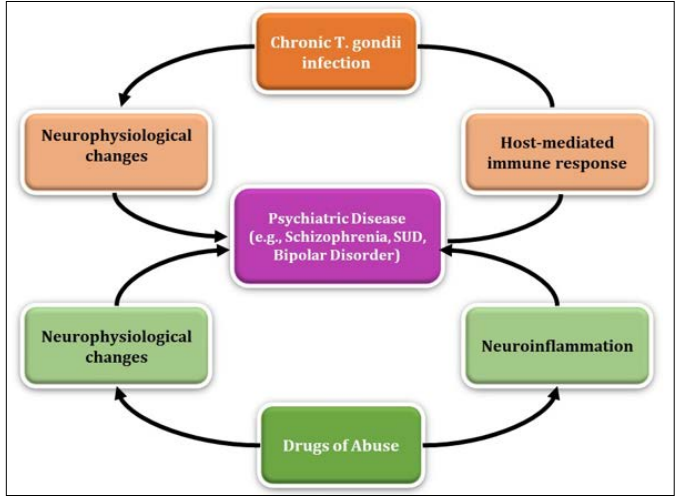
Figure 5: Drugs of abuse and chronic T. gondii infection display parallel mechanisms when acting on the brain that may be implicated in the development of psychiatric disease
Alcohol exposure can cause dysregulation of the immune system; however, the association between T. gondii infection and alcohol consumption is not well understood. In a study looking at the seroepidemiology of infection with T. gondii in Durango, Mexico, there was a positive association between alcohol consumption and anti-T. gondii IgG and IgM antibodies, specifically an 11.2% seroprevalence [93]. Additionally, those infected with T. gondii have displayed increased risky behavior due to the parasite’s ability to infect and persist in the central nervous system. Previous studies have attributed risky behavior to increased road traffic accidents and suicidal behaviors [94]. Samojlowicz et al studied this association between latent toxoplasmosis and excessive alcohol consumption by observing seroprevalence in three groups: risky behavior, control, and inconclusively risky behavior [95]. The risky behavior group included data from deaths attributed to risky behavior such as: traffic accidents, suicide, substance overdose, and other risky behavior, such as disregard for reasonable safety precautions. Of the 535 individuals used in the study, anti IgG antibodies were found in 46.4% of all evaluated, with 51.1% of seropositive individuals being in the risky behavior group. When observing individual causes of death in the risky behavior group, a higher proportion of seropositive subjects were associated with substance overdose (53%, with 29.9% attributed to alcohol overdose) and other risky behavior (55%). Ultimately, the data indicated a strong positive correlation between anti- T. gondii IgG antibodies and alcohol consumption when considering the relationship between T. gondii infection and risky behavior [95]. The underlying mechanism of parasite-induced behavior modification in humans is still unclear; however, evidence suggests that the presence of cysts diffusely localized throughout the brain plays a major role. One hypothesis for T gondii’s ability to manipulate behavior involves the dysregulation of dopaminergic (DA) signaling in the brain, specifically in the nucleus accumbens (NAc) and ventral tegmental area (VTA). Moreover, infected rats display reduced DA content in the NAc and an increase in impulsivity compared to controls [96]. In addition, Berdoy et al demonstrated an increase in attraction rather than aversion when T. gondii infected rodents were exposed to odors from the members of the family Felidae, suggesting that the parasite alters neurological activity of its intermediate hosts [97]. Regarding other areas of the brain, Gatkowska et al explained that the presence of cysts in the hippocampus and amygdala revealed diminished exploratory activity and less grooming behavior in infected mice compared to uninfected mice [98].
Given that the NAc and VTA are areas of the brain important for regulating reward and reinforcement, there is a well described association between alcohol and dopamine transmission in the NAc and VTA [99]. Doyon et al observed DA neurons being stimulated by alcohol and an increase in the proportion of these neurons exhibiting pacemaker-like firing patterns [100]. In addition to its role in the brain, alcohol has been shown to significantly alter immune function and inflammation in those affected. When administered daily doses of alcohol, mice exhibited an increase in proinflammatory cytokines, TNF-α, and MCP-I, as well as an enhanced cytokine response when pretreating lipopolysaccharide (LPS) induced mice with alcohol [101]. More importantly, a prolonged proinflammatory response and subsequent decrease in IL-10 production in the brain occurs, as opposed to occurring in the liver and serum of alcohol exposed mice [101]. Taken together, these studies suggest an increased risk of T. gondii infection for those who consume or are exposed to alcohol. However, further research must be conducted to understand the specific cytokines expressed when simultaneously exposed to alcohol and infected with T. gondii in both the acute and chronic stages.
T.gondii infection has been linked to several behavioral and psychiatric disorders due to its presence in the brain and influence on dopamine transmission. For example, T. gondii seropositivity has been associated with schizophrenia due to abnormal dopamine transmission as well as a risk factor for the development of depression and other psychiatric disorders [102,103]. In addition to dopamine, there was a significantly higher prevalence of anti- T. gondii IgG antibodies among patients in Western Romania with schizophrenia (69.77%), personality/behavior disorders (76.74%), and mental disorders concerning alcohol abuse (84.62%) compared to controls (p = 0.009, p = 0.005, p = 0.043, respectively) [104]. Likewise, substance use is linked to the manipulation of dopamine transmission and the development of these diseases [105]. But there is still a lack of information associated with T. gondii infection and substance use. In a case control study performed in 2015 by Alvarado-Esquivel et al, it appeared that behaviors associated with substance abuse did not increase the risk T. gondii infection, suggesting that latent toxoplasmosis may influence the degree of substance use rather than substance use increasing likelihood of infection [106]. Additionally, in a case control study conducted by Sharifzadeh et al, drug dependent individuals in Iran were shown to have higher exposure to T. gondii, through seroprevalence of anti-T. gondii IgG antibody, compared to non-dependent individuals. The study further discussed the socioeconomic impact on drug dependent individuals as a contributing factor toward exposure to T. gondii infection [107]. Moreover, Berrett et al observed that T. gondii seropositivity was associated with a reduced likelihood of self-reported substance use, e.g., tobacco, marijuana, heroin, and methamphetamines. The study discusses that this reduction in self-reporting could be due to T. gondii’s influence on dopaminergic signaling in the brain [108]. This data illustrates an association between T. gondii and substance use, particularly with the overlap of dopaminergic signaling in both the parasite’s life cycle and drug dependence. Further research must be conducted to fully understand the biological effects of these associations.
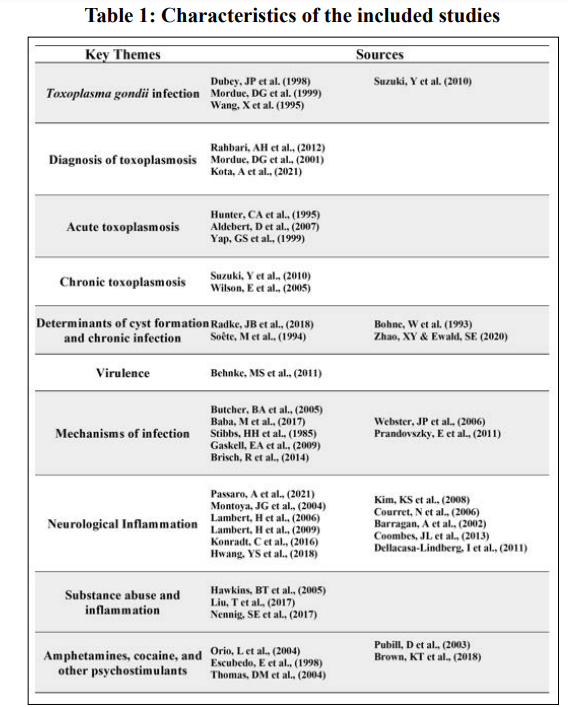
There is still ongoing research into the inflammatory effects of specific drugs of abuse, with preliminary data showing mixed results. As stated previously, some drugs of abuse have immunosuppressive effects, such as alcohol, posing the question of whether those with SUD may protect T. gondii from host immunity during chronic infection. The highly stimulating effects of these substances in the brain and their ability to disrupt the BBB permeability pose a risk for further neurological damage associated with T. gondii infection. Currently, research on the immunological effects of alcohol is more prevalent compared to other drugs of abuse, making alcohol abuse a useful model for understanding T. gondii infection in the context of drug dependence. In the Durango, Mexico seroepidemiology study, the positive association between alcohol consumption and anti- T. gondii antibodies provides preliminary evidence of the link between the two. However, it may be worth simplifying the research to understand the key biological markers and cytokine expression involved in T. gondii infection under alcohol exposure. This may elucidate the mechanisms of parasite entry and the potential protective effects employed by alcohol for T. gondii to evade host immunity. Understanding these mechanisms may provide foundational knowledge of this complex parasite’s effects in the brain and the additional risks of SUD in the context of opportunistic infections (Table 1).
Aside from the immunological perspective, chronic T. gondii infection can lead to behavioral changes such as impulsivity, aggression, and self-directed violence. Additionally, T. gondii infection has been linked to the development of schizophrenia and other psychiatric diseases due to its increase in dopamine metabolism, neuromodulating effects, and the presence of tyrosine hydroxylase in its genome. The studies evaluating dopamine antagonist medication, such as haloperidol, on T. gondii associated behavior changes could be expanded into therapeutic use for
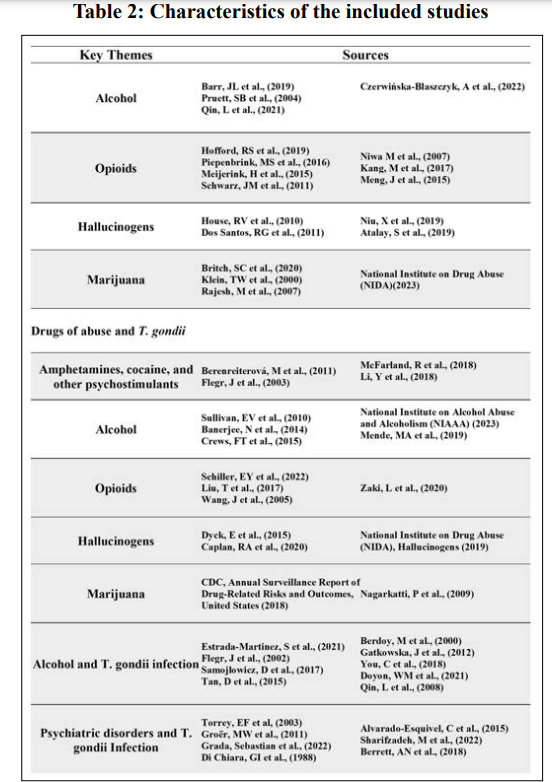
The parallels between these entities poses two questions:1) whether T. gondii infection can predispose those with an SUD to developing further psychiatric disease and 2) whether SUD is a risk factor for developing behavioral and psychiatric complications associated with T. gondii infection (Table 2).
The current state of research on T. gondii infection is expanding, but there remain several unanswered questions regarding its inflammatory response and neuropsychiatric consequences. Likewise, research on drugs of abuse have long been untouched, but with the recent support, attempts to understand their effects have increased. The purpose of this review was to provide a comprehensive summary of the existing research on both topics and the potential connections between the two. The current limitations in research include a gap in knowledge on T. gondii’s cytokine expression profile upon parasite entry to the brain, its effects on the inflammatory response, and dopamine metabolism of T. gondii infection upon administration of drugs of abuse. Additionally, further research is necessary to understand the immunomodulating effects of various drugs of abuse in the context of T. gondii infection, thus providing foundational knowledge on potential protective mechanisms. Limitations of this review include excluded literature due to narrowed analysis of studies found under specific keywords in each database. Based on the results, this review provides foundational knowledge on the hypothesis that SUD is a potential risk factor for behavioral and psychiatric complications associated with T. gondii infection. However, further research is necessary to understand T. gondii infection in the context of drug dependence, withdrawal, and the complex development of SUD.
Not applicable
The author(s) declare that there is no conflict of interest regarding the publication of this paper.
This research received no external funding.
An Acknowledgements section is optional and may recognise those individuals who provided help during the research and preparation of the manuscript.
PRISMA-ScR Checklist
AS provided substantial contributions to conception and design, acquisition of data, or analysis and interpretation of data; AS and VJ drafted the article or revised it critically for important intellectual content; VJ gave final approval of the version of the article to be published; all authors agree to be accountable for all aspects of the work in ensuring that questions related to the accuracy or integrity of any part of the work are appropriately investigated and resolved. All authors have read and agreed to the published version of the manuscript.